2,281 posters, 63 videos, 56 audios, 43 topics, 18 sessions, 6,174 authors, 2,323 institutions
ePostersLive by SciGen Technologies S.A. All rights reserved.
RCOG World Congress 2025
June 23 - 25, 2025 | ExCeL London, UK
EP.0382
Surgical Management of Grade IV Deeply Infiltrating Endometriosis with Gastrointestinal Involvement
Poster Presenter
Part of Topic
Endometriosis & uterine disorders
Video
Title: Surgical Management of Grade IV Deeply Infiltrating Endometriosis with Gastrointestinal Involvement: A Case Report
Objective:
Endometriosis is a relatively common condition; however, deeply infiltrating endometriosis (DIE) with the involvement of the GI system is less common. The prevalence of intestinal endometriosis has been estimated to be between 3% and 37% of all endometriosis cases. Therefore, providing effective surgical treatment for those cases can be challenging and require multidisciplinary experiments. This abstract presents a case report and surgical video of a patient undergoing excision of grade IV infiltrating endometriosis, with a focus on maximizing fertility potential and preserving bowel function.
Case report:
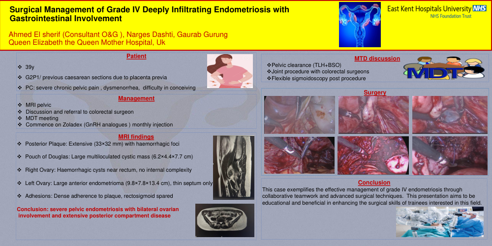
We present the case of a 39-year-old woman, G2P1, who was referred by her general practitioner to gynaecology due to difficulty conceiving. She also had a history of previous caesarean sections due to placenta previa.
Following investigations, including ultrasound and MRI, Severe endometriosis was diagnosed, showing extensive involvement of the middle and posterior compartments. Key findings included:
A large endometrioma involving the left ovary,
A cystic mass with a haemorrhagic component in the pouch of Douglas, arising from the right adnexa
A plaque on the posterior aspect of the uterus
A plaque on the anterior wall of the rectum
Discussion
The case was reviewed in a MTD meeting, which included colorectal surgeons to develop a comprehensive care plan. Preoperatively, the patient was started on monthly Zoledex injections for six months to reduce pelvic inflammation.
She underwent laparoscopic excision of endometriosis, including adhesiolysis of the endometrioma. Techniques were employed to maximise her future fertility potential by preserving both ovaries. During the procedure, the rectosigmoid was successfully separated from the endometriotic plaque attached to the posterior uterine wall, and there was no significant involvement of the rectosigmoid, hence bowel resection was unnecessary. At the conclusion of the procedure, there were no signs of injury, and the bowel appeared healthy on the rigid sigmoidoscopy. The plaques on the uterus and vagina were excised without complications. Also, a small endometrioma on the posterior vaginal wall was drained and sutured laparoscopically.
Postoperatively, the patient continued monthly GnRH analog treatment to manage any residual disease and prevent flare-ups until fertility was desired. She was informed about the potential risks of long-term GnRH analog use, including the need for regular bone density monitoring.
Conclusions:
This case exemplifies the effective management of grade IV endometriosis through collaborative teamwork and advanced surgical techniques. This presentation aims to be educational and beneficial in enhancing the surgical skills of trainees interested in this field.