2,281 posters, 63 videos, 56 audios, 43 topics, 18 sessions, 6,174 authors, 2,323 institutions
ePostersLive by SciGen Technologies S.A. All rights reserved.
RCOG World Congress 2025
June 23 - 25, 2025 | ExCeL London, UK
EP.0371
A case of Unicornuate uterus with hydrosalpinx
Poster Presenter
Authors
Reshma Vellaparambil Parameswaran Nair, Abdul rahman Al farsi
Affiliations
Part of Topic
Endometriosis & uterine disorders
A CASE OF UNICORNUATE UTERUS WITH HYDROSALPINX
Dr. Reshma Vellaparambil Parameswaran Nair & Dr. Abdul Rahman Al Farsi
INTRODUCTION
Congenital uterine anomalies are deviations from normal anatomy occurring as a result of defective development of Müllerian duct during embryological period. Most of such anomalies except some are asymptomatic. Defects in development of Müllerian ducts leads to agenesis or hypoplasia resulting in Unicornuate uterus. Unification defects such as unicornuate uterus do not reduce fertility but increase risk of first trimester miscarriage, fetal malpresentation and preterm labour. Hydrosalpinx is fluid filled distended fallopian tube and pelvic infections are one of the reason for its occurrence.
CASE SUMMARY
A 32-year-old Nulliparous lady, married 7 years started planning pregnancy few months back presented with pain lower abdomen, delayed periods, spotting per vaginum and a positive pregnancy test. Her Beta Hcg at that time was found to be 235, ultrasound scan was showing left ectopic pregnancy. Patient was managed medically with methotrexate and her pregnancy resolved. After 6 months following this ectopic pregnancy she presented with delayed periods, negative pregnancy test. Ultrasound showed polycystic ovaries.
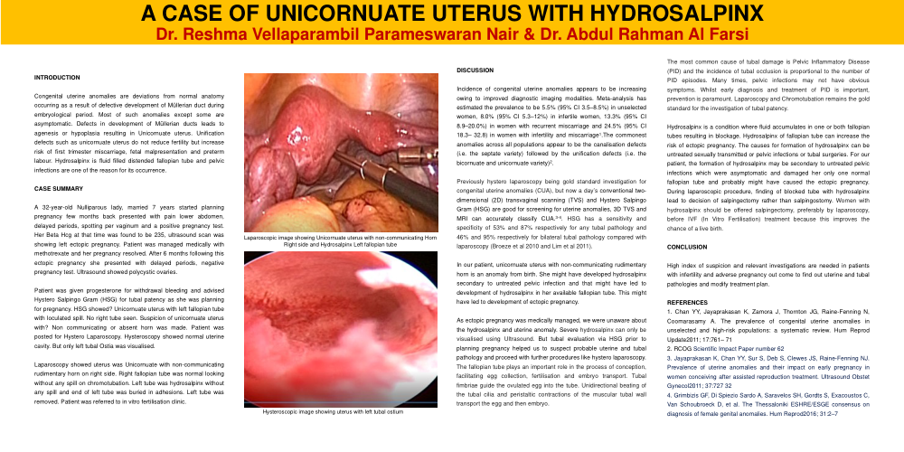
Patient was given progesterone for withdrawal bleeding and advised Hystero Salpingo Gram (HSG) for tubal patency as she was planning for pregnancy. HSG showed? Unicornuate uterus with left fallopian tube with loculated spill. No right tube seen. Suspicion of unicornuate uterus with? Non communicating or absent horn was made. Patient was posted for Hystero Laparoscopy. Hysteroscopy showed normal uterine cavity. But only left tubal Ostia was visualised.
Laparoscopy showed uterus was Unicornuate with non-communicating rudimentary horn on right side. Right fallopian tube was normal looking without any spill on chromotubation. Left tube was hydrosalpinx without any spill and end of left tube was buried in adhesions. Left tube was removed. Patient was referred to in vitro fertilisation clinic.
DISCUSSION
Incidence of congenital uterine anomalies appears to be increasing owing to improved diagnostic imaging modalities. Meta-analysis has estimated the prevalence to be 5.5% (95% CI 3.5–8.5%) in unselected women, 8.0% (95% CI 5.3–12%) in infertile women, 13.3% (95% CI 8.9–20.0%) in women with recurrent miscarriage and 24.5% (95% CI 18.3– 32.8) in women with infertility and miscarriage1.The commonest anomalies across all populations appear to be the canalisation defects (i.e. the septate variety) followed by the unification defects (i.e. the bicornuate and unicornuate variety)2.
Previously hystero laparoscopy being gold standard investigation for congenital uterine anomalies (CUA), but now a day’s conventional two-dimensional (2D) transvaginal scanning (TVS) and Hystero Salpingo Gram (HSG) are good for screening for uterine anomalies, 3D TVS and MRI can accurately classify CUA.3-4. HSG has a sensitivity and specificity of 53% and 87% respectively for any tubal pathology and 46% and 95% respectively for bilateral tubal pathology compared with laparoscopy (Broeze et al 2010 and Lim et al 2011).
In our patient, unicornuate uterus with non-communicating rudimentary horn is an anomaly from birth. She might have developed hydrosalpinx secondary to untreated pelvic infection and that might have led to development of hydrosalpinx in her available fallopian tube. This might have led to development of ectopic pregnancy.
As ectopic pregnancy was medically managed, we were unaware about the hydrosalpinx and uterine anomaly. Severe hydrosalpinx can only be visualised using Ultrasound. But tubal evaluation via HSG prior to planning pregnancy helped us to suspect probable uterine and tubal pathology and proceed with further procedures like hystero laparoscopy. The fallopian tube plays an important role in the process of conception, facilitating egg collection, fertilisation and embryo transport. Tubal fimbriae guide the ovulated egg into the tube. Unidirectional beating of the tubal cilia and peristaltic contractions of the muscular tubal wall transport the egg and then embryo.
The most common cause of tubal damage is Pelvic Inflammatory Disease (PID) and the incidence of tubal occlusion is proportional to the number of PID episodes. Many times, pelvic infections may not have obvious symptoms. Whilst early diagnosis and treatment of PID is important, prevention is paramount. Laparoscopy and Chromotubation remains the gold standard for the investigation of tubal patency.
Hydrosalpinx is a condition where fluid accumulates in one or both fallopian tubes resulting in blockage. Hydrosalpinx of fallopian tube can increase the risk of ectopic pregnancy. The causes for formation of hydrosalpinx can be untreated sexually transmitted or pelvic infections or tubal surgeries. For our patient, the formation of hydrosalpinx may be secondary to untreated pelvic infections which were asymptomatic and damaged her only one normal fallopian tube and probably might have caused the ectopic pregnancy. During laparoscopic procedure, finding of blocked tube with hydrosalpinx lead to decision of salpingectomy rather than salpingostomy. Women with hydrosalpinx should be offered salpingectomy, preferably by laparoscopy, before IVF (In Vitro Fertilisation) treatment because this improves the chance of a live birth.
CONCLUSION
High index of suspicion and relevant investigations are needed in patients with infertility and adverse pregnancy out come to find out uterine and tubal pathologies and modify treatment plan.
REFERENCES
1. Chan YY, Jayaprakasan K, Zamora J, Thornton JG, Raine-Fenning N, Coomarasamy A. The prevalence of congenital uterine anomalies in unselected and high-risk populations: a systematic review. Hum Reprod Update2011; 17:761– 71
2. RCOG Scientific Impact Paper number 62
3. Jayaprakasan K, Chan YY, Sur S, Deb S, Clewes JS, Raine-Fenning NJ. Prevalence of uterine anomalies and their impact on early pregnancy in women conceiving after assisted reproduction treatment. Ultrasound Obstet Gynecol2011; 37:727 32
4. Grimbizis GF, Di Spiezio Sardo A, Saravelos SH, Gordts S, Exacoustos C, Van Schoubroeck D, et al. The Thessaloniki ESHRE/ESGE consensus on diagnosis of female genital anomalies. Hum Reprod2016; 31:2–7