2,281 posters, 63 videos, 56 audios, 43 topics, 18 sessions, 6,174 authors, 2,323 institutions
ePostersLive by SciGen Technologies S.A. All rights reserved.
RCOG World Congress 2025
June 23 - 25, 2025 | ExCeL London, UK
EP.0354
Application of Indocyanine Green and Near Infrared Fluorescence Imaging in endometriosis management
Poster Presenter
Authors
Elias Tsakos, Emmanouil M. Xydias, Nikolaos Tsagias, Nikolaos Plevris, Alexandros Daponte, Apostolos Ziogas, Kanelina Bimpa
Affiliations
EmbryoClinic IVF, University of Thessaly, School of Health Sciences, Faculty of Medicine, Metropolitan General Hospital, 1st Gynaecological Department, Division of Robotic and Laparoscopic Surgery
Part of Topic
Endometriosis & uterine disorders
Application of Indocyanine Green and Near Infrared Fluorescence Imaging in endometriosis management
Dr. Elias Tsakos1, Dr. Emmanouil M. Xydias1, Dr. Nikolaos Tsagias1, Dr. Nikolaos Plevris2, Prof. Alexandros Daponte3, Dr. Apostolos Ziogas3, Dr. Kanelina Bimpa1
1EmbryoClinic IVF, Thessaloniki, Greece. 2Metropolitan General Hospital, 1st Gynaecological Department, Division of Robotic and Laparoscopic Surgery, Athens, Greece. 3University of Thessaly, School of Health Sciences, Faculty of Medicine, Larissa, Greece
Introduction
Near infrared fluorescence (NIRF) imaging with the use of Indocyanine Green (ICG) has been widely used in the field of Gynaecological Oncology for lymphnode mapping with remarkable success. A novel, highly promising application of this modality with significant potential is in the identification of endometriosis lesions and in guiding their management. The aim of this review and meta-analysis is to examine the different applications of NIRF-ICG in the modern surgical treatment of endometriosis.
Methods
A systematic literature search was performed in the Scopus, Web of science and PubMed databases in accordance with the PRISMA 2020 guidelines. Studies were evaluated for relevance and included in the qualitative synthesis. Where sufficient data were available, meta-analysis was also performed.
Results
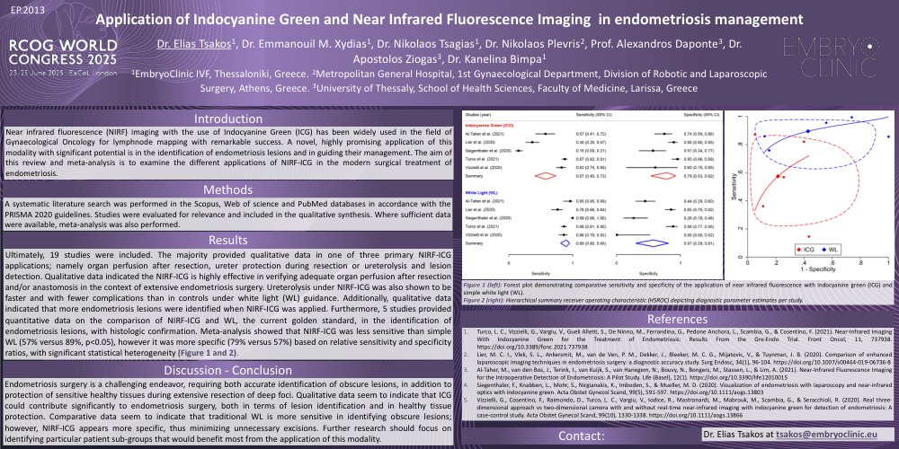
Ultimately, 19 studies were included. The majority provided qualitative data in one of three primary NIRF-ICG applications; namely organ perfusion after resection, ureter protection during resection or ureterolysis and lesion detection. Qualitative data indicated the NIRF-ICG is highly effective in verifying adequate organ perfusion after resection and/or anastomosis in the context of extensive endometriosis surgery. Ureterolysis under NIRF-ICG was also shown to be faster and with fewer complications than in controls under white light (WL) guidance. Additionally, qualitative data indicated that more endometriosis lesions were identified when NIRF-ICG was applied. Furthermore, 5 studies provided quantitative data on the comparison of NIRF-ICG and WL, the current golden standard, in the identification of endometriosis lesions, with histologic confirmation. Meta-analysis showed that NIRF-ICG was less sensitive than simple WL (57% versus 89%, p<0.05), however it was more specific (79% versus 57%) based on relative sensitivity and specificity ratios, with significant statistical heterogeneity (Figure 1 and 2).
Discussion - Conclusion
Endometriosis surgery is a challenging endeavor, requiring both accurate identification of obscure lesions, in addition to protection of sensitive healthy tissues during extensive resection of deep foci. Qualitative data seem to indicate that ICG could contribute significantly to endometriosis surgery, both in terms of lesion identification and in healthy tissue protection. Comparative data seem to indicate that traditional WL is more sensitive in identifying obscure lesions; however, NIRF-ICG appears more specific, thus minimizing unnecessary excisions. Further research should focus on identifying particular patient sub-groups that would benefit most from the application of this modality.
References
Dr. Elias Tsakos at tsakos@embryoclinic.eu
Figure 1 (left): Forest plot demonstrating comparative sensitivity and specificity of the application of near infrared fluorescence with indocyanine green (ICG) and simple white light (WL).
Figure 2 (right): Hierarchical summary receiver operating characteristic (HSROC) depicting diagnostic parameter estimates per study.