2,281 posters, 63 videos, 56 audios, 43 topics, 18 sessions, 6,174 authors, 2,323 institutions
ePostersLive by SciGen Technologies S.A. All rights reserved.
RCOG World Congress 2025
June 23 - 25, 2025 | ExCeL London, UK
EP.2094
A pilot study of a Cu-IUD insertion technique (PPIUD) at caesarean section
Poster Presenter
Authors
Affiliations
Part of Topic
Sexual health & contraception/sexual health & HIV
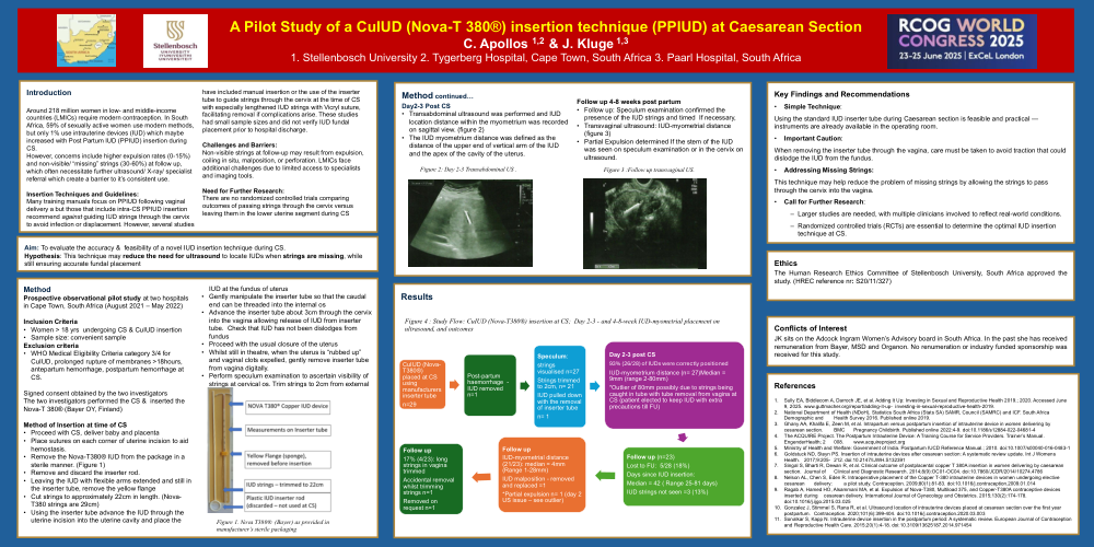
A Pilot Study of a CuIUD (Nova-T 380®) insertion technique (PPIUD) at Caesarean Section
C. Apollos 1,2 & J. Kluge 1,3
1. Stellenbosch University 2. Tygerberg Hospital, Cape Town, South Africa 3. Paarl Hospital, South Africa
Introduction
Around 218 million women in low- and middle-income countries (LMICs) require modern contraception. In South Africa, 59% of sexually active women use modern methods, but only 1% use intrauterine devices (IUD) which maybe increased with Post Partum IUD (PPIUD) insertion during CS.
However, concerns include higher expulsion rates (0-15%) and non-visible/ “missing” strings (30-60%) at follow up, which often necessitate further ultrasound/ X-ray/ specialist referral which create a barrier to it’s consistent use.
Insertion Techniques and Guidelines:
Many training manuals focus on PPIUD following vaginal delivery a but those that include intra-CS PPIUD insertion recommend against guiding IUD strings through the cervix to avoid infection or displacement. However, several studies have included manual insertion or the use of the inserter tube to guide strings through the cervix at the time of CS with especially lengthened IUD strings with Vicryl suture, facilitating removal if complications arise. These studies had small sample sizes and did not verify IUD fundal placement prior to hospital discharge.
Challenges and Barriers:
Non-visible strings at follow-up may result from expulsion, coiling in situ, malposition, or perforation. LMICs face additional challenges due to limited access to specialists and imaging tools.
Need for Further Research:
There are no randomized controlled trials comparing outcomes of passing strings through the cervix versus leaving them in the lower uterine segment during CS
Aim: To evaluate the accuracy & feasibility of a novel IUD insertion technique during CS.
Hypothesis: This technique may reduce the need for ultrasound to locate IUDs when strings are missing, while still ensuring accurate fundal placement
Method
Prospective observational pilot study at two hospitals in Cape Town, South Africa (August 2021 – May 2022)
Inclusion Criteria
Exclusion criteria
Signed consent obtained by the two investigators
The two investigators performed the CS & inserted the Nova-T 380® (Bayer OY, Finland)
Method of Insertion at time of CS
Perform speculum examination to ascertain visibility of strings at cervical os. Trim strings to 2cm from external
Method continued…
Day2-3 Post CS
Follow up 4-8 weeks post partum
Post-partum haemorrhage occurred in one participant and the IUD was removed. Ninety-three percent (26/28) of IUDs were adequately positioned on day 2-3 post-delivery with the median IUD -myometrial distance of 9mm (range 2- 80mm).
Key Findings and Recommendations
• Simple Technique:
Using the standard IUD inserter tube during Caesarean section is feasible and practical — instruments are already available in the operating room.
• Important Caution:
When removing the inserter tube through the vagina, care must be taken to avoid traction that could dislodge the IUD from the fundus.
• Addressing Missing Strings:
This technique may help reduce the problem of missing strings by allowing the strings to pass through the cervix into the vagina.
• Call for Further Research:
Larger studies are needed, with multiple clinicians involved to reflect real-world conditions.
Randomized controlled trials (RCTs) are essential to determine the optimal IUD insertion technique at CS.
Ethics
The Human Research Ethics Committee of Stellenbosch University, South Africa approved the study. (HREC reference nr: S20/11/327)
Conflicts of Interest
JK sits on the Adcock Ingram Women’s Advisory board in South Africa. In the past she has received remuneration from Bayer, MSD and Organon. No renumeration or industry funded sponsorship was received for this study.
References
1. Sully EA, Biddlecom A, Darroch JE, et al. Adding It Up: Investing in Sexual and Reproductive Health 2019.; 2020. Accessed June 9, 2025. www.guttmacher.org/report/adding-it-up- investing-in-sexual-reproductive-health-2019.
2. National Department of Health (NDoH), Statistics South Africa (Stats SA) SAMR, Council (SAMRC) and ICF. South Africa Demographic and Health Survey 2016. Published online 2019.
3. Ghany AA, Khalifa E, Zeen M, et al. Intrapartum versus postpartum insertion of intrauterine device in women delivering by cesarean section. BMC Pregnancy Childbirth. Published online 2022:4-9. doi:10.1186/s12884-022-04681-4
4. The ACQUIRE Project. The Postpartum Intrauterine Device: A Training Course for Service Providers. Trainer’s Manual. EngenderHealth; 2 008. www.acquireproject.org
5. Ministry of Health and Welfare: Government of India. Postpartum IUCD Reference Manual.; 2010. doi:10.1007/s00040-016-0483-1
6. Goldstuck ND, Steyn PS. Insertion of intrauterine devices after cesarean section: A systematic review update. Int J Womens Health. 2017;9:205- 212. doi:10.2147/IJWH.S132391
7. Singal S, Bharti R, Dewan R, et al. Clinical outcome of postplacental copper T 380A insertion in women delivering by caesarean section. Journal of Clinical and Diagnostic Research. 2014;8(9):OC01-OC04. doi:10.7860/JCDR/2014/10274.4786
8. Nelson AL, Chen S, Eden R. Intraoperative placement of the Copper T-380 intrauterine devices in women undergoing elective cesarean delivery: a pilot study. Contraception. 2009;80(1):81-83. doi:10.1016/j.contraception.2009.01.014
9. Ragab A, Hamed HO, Alsammani MA, et al. Expulsion of Nova-T380, Multiload 375, and Copper-T380A contraceptive devices inserted during cesarean delivery. International Journal of Gynecology and Obstetrics. 2015;130(2):174-178. doi:10.1016/j.ijgo.2015.03.025
10. Gonzalez J, Stimmel S, Rana R, et al. Ultrasound location of intrauterine devices placed at cesarean section over the first year postpartum. Contraception. 2020;101(6):399-404. doi:10.1016/j.contraception.2020.03.003
11. Sonalkar S, Kapp N. Intrauterine device insertion in the postpartum period: A systematic review. European Journal of Contraception and Reproductive Health Care. 2015;20(1):4-18. doi:10.3109/13625187.2014.971454