2,281 posters, 63 videos, 56 audios, 43 topics, 18 sessions, 6,174 authors, 2,323 institutions
ePostersLive by SciGen Technologies S.A. All rights reserved.
RCOG World Congress 2025
June 23 - 25, 2025 | ExCeL London, UK
EP.1679
Case report: An unusual case of a thyroid mass in pregnancy
Poster Presenter
Authors
Affiliations
Part of Topic
Maternal medicine
Case report: An unusual case of a thyroid mass in pregnancy
N. Haringman, M Tiwara, G Lysycia, A Govind
North Middlesex University Hospital
We describe an unusual presentation of pregnancy related thyroid cancer, diagnosed antenatally, subsequent to a presentation with symptoms of airway obstruction.
Current and recent pregnancy has been found to be associated with a transient increased risk of thyroid cancer (2,3,4). As well as increasing the risk of thyroid cancer, pregnancy also affects the treatment plan (5), which needs to be tailored to the patient.
It is unclear whether pregnancy affects the prognosis in patients with thyroid cancer. Some studies indicated no significant effect on prognosis (6), however another one suggested that pregnancy increases the risk of persistent and recurrent disease (7).
A 20 year old patient presented to hospital at 36+1 weeks gestation with a one month history of a cough and increasing shortness of breath. Her risk factors at booking include being a late booker, a first cousin consanguineous marriage, and limited English. She had booked late for antenatal care due to being abroad in Turkey until 24 weeks gestation. She had had normal ultrasound scans, with an uneventful antenatal course prior to her presentation.
At 36 weeks gestation, she reported a thyroid mass, and was sent to maternity triage, following an antenatal clinic appointment. She reported that she had noticed this for several months, however she had not previously disclosed this during her prior appointments.
On examination her chest was clear, however she had stridor, and a large thyroid mass causing tracheal deviation. A hoarse voice was also noted. She denied any history of thyroid issues, or infectious symptoms.
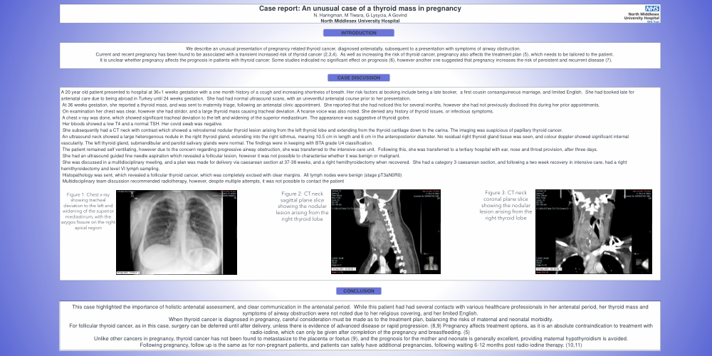
A chest x-ray was done, which showed significant tracheal deviation to the left and widening of the superior mediastinum. The appearance was suggestive of thyroid goitre.
Her bloods showed a low T4 and a normal TSH. Her covid swab was negative.
She subsequently had a CT neck with contrast which showed a retrosternal nodular thyroid lesion arising from the left thyroid lobe and extending from the thyroid cartilage down to the carina. The imaging was suspicious of papillary thyroid cancer.
An ultrasound neck showed a large heterogenous nodule in the right thyroid gland, extending into the right isthmus, mearing 10.5 cm in length and 6 cm in the anteroposterior diameter. No residual right thyroid gland tissue was seen, and colour doppler showed significant internal vascularity. The left thyroid gland, submandibular and parotid salivary glands were normal. The findings were in keeping with BTA grade U4 classification.
The patient remained self ventilating, however due to the concern regarding progressive airway obstruction, she was transferred to the intensive care unit. Following this, she was transferred to a tertiary hospital with ear, nose and throat provision, after three days.
She had an ultrasound guided fine needle aspiration which revealed a follicular lesion, however it was not possible to characterise whether it was benign or malignant.
She was discussed in a multidisciplinary meeting, and a plan was made for delivery via caesarean section at 37-38 weeks, and a right hemithyroidectomy when recovered. She had a category 3 caesarean section, and following a two week recovery in intensive care, had a right hemithyroidectomy and level VI lymph sampling.
Histopathology was sent, which revealed a follicular thyroid cancer, which was completely excised with clear margins. All lymph nodes were benign (stage pT3aN0R0)
Multidisciplinary team discussion recommended radiotherapy, however, despite multiple attempts, it was not possible to contact the patient.
This case highlighted the importance of holistic antenatal assessment, and clear communication in the antenatal period. While this patient had had several contacts with various healthcare professionals in her antenatal period, her thyroid mass and symptoms of airway obstruction were not noted due to her religious covering, and her limited English.
When thyroid cancer is diagnosed in pregnancy, careful consideration must be made as to the treatment plan, balancing the risks of maternal and neonatal morbidity.
For follicular thyroid cancer, as in this case, surgery can be deferred until after delivery, unless there is evidence of advanced disease or rapid progression. (8,9) Pregnancy affects treatment options, as it is an absolute contraindication to treatment with radio-iodine, which can only be given after completion of the pregnancy and breastfeeding. (5)
Unlike other cancers in pregnancy, thyroid cancer has not been found to metastasize to the placenta or foetus (9), and the prognosis for the mother and neonate is generally excellent, providing maternal hypothyroidism is avoided.
Following pregnancy, follow up is the same as for non-pregnant patients, and patients can safely have additional pregnancies, following waiting 6-12 months post radio-iodine therapy. (10,11)