2,281 posters, 63 videos, 56 audios, 43 topics, 18 sessions, 6,174 authors, 2,323 institutions
ePostersLive by SciGen Technologies S.A. All rights reserved.
RCOG World Congress 2025
June 23 - 25, 2025 | ExCeL London, UK
EP.1670
A rare case of Lymphangioleiomyomatosis (LAM) in pregnancy
Poster Presenter
Part of Topic
Maternal medicine
A rare case of lymphangioleiomyomatosis (LAM) in pregnancy
M Malina, E Phillips, M Dissanayake, J O’Heney, K Wiles, S Sivarajasingam
Background and Objective
•Lymphangioleiomyomatosis (LAM) is a rare systemic condition (4 in 1,000,000).
•LAM is oestrogen driven affecting almost exclusively reproductive age females.
•Progressive cystic destruction of the lungs occurs.
•Risks in pregnancy include deteriorating lung function, spontaneous pneumo-thorax and preterm birth.
•We report a successful pregnancy outcome in a primigravida with LAM who had a Caesarean birth at 36 weeks’ gestation.
•Care was managed at a local hospital in East London with the support of the North-East London Maternal Medicine Network.
Case Report
•31-year-old, no previous pregnancies
•LAM diagnosed 3 years prior to pregnancy after a spontaneous pneumothorax
•Under specialist care at the UK national LAM centre.
•Pre-pregnancy management included a left-sided bullectomy and pleurodesis, and maintenance sirolimus.
•Pre-pregnancy FEV1 and FVC 50-80% predicted.
•
Pregnancy care (see fig 1):
•Sirolimus discontinued.
•Respiratory infection in first trimester with wheeze managed with oral and inhaled corticosteroids.
•Echocardiogram: normal LV and RV function.
•Cervical length monitoring for previous large loop excision of the transformation zone (LLETZ).
•Borderline shortened cervix managed with vaginal progesterone.
•Regular obstetric medicine review.
•Fetal medicine scanning.
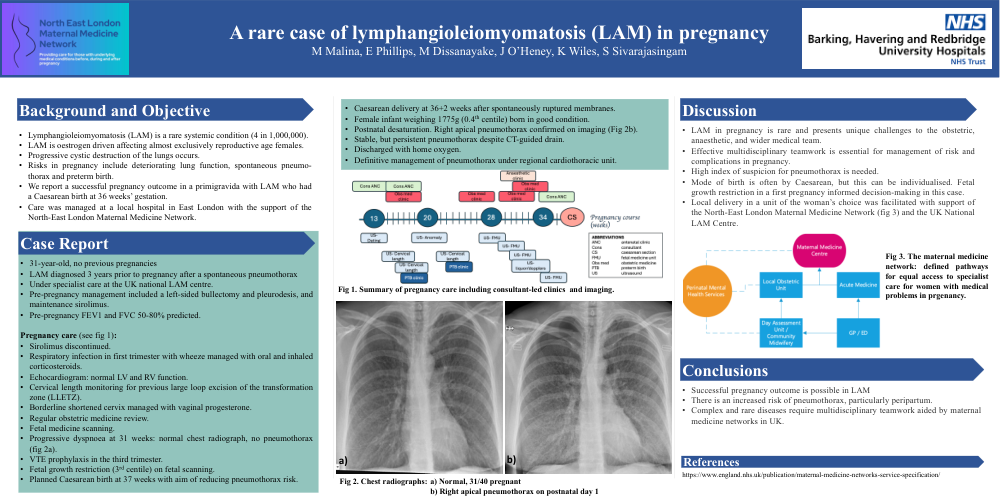
•Progressive dyspnoea at 31 weeks: normal chest radiograph, no pneumothorax (fig 2a).
•VTE prophylaxis in the third trimester.
•Fetal growth restriction (3rd centile) on fetal scanning.
•Planned Caesarean birth at 37 weeks with aim of reducing pneumothorax risk.
•Caesarean delivery at 36+2 weeks after spontaneously ruptured membranes.
•Female infant weighing 1775g (0.4th centile) born in good condition.
•Postnatal desaturation. Right apical pneumothorax confirmed on imaging (Fig 2b).
•Stable, but persistent pneumothorax despite CT-guided drain.
•Discharged with home oxygen.
•Definitive management of pneumothorax under regional cardiothoracic unit.
Discussion
•LAM in pregnancy is rare and presents unique challenges to the obstetric, anaesthetic, and wider medical team.
•Effective multidisciplinary teamwork is essential for management of risk and complications in pregnancy.
•High index of suspicion for pneumothorax is needed.
•Mode of birth is often by Caesarean, but this can be individualised. Fetal growth restriction in a first pregnancy informed decision-making in this case.
•Local delivery in a unit of the woman’s choice was facilitated with support of the North-East London Maternal Medicine Network (fig 3) and the UK National LAM Centre.
Conclusions
•Successful pregnancy outcome is possible in LAM
•There is an increased risk of pneumothorax, particularly peripartum.
•Complex and rare diseases require multidisciplinary teamwork aided by maternal medicine networks in UK.