Loading...

2,281 posters, 63 videos, 56 audios, 43 topics, 18 sessions, 6,174 authors, 2,323 institutions

ePostersLive by SciGen Technologies S.A. All rights reserved.

RCOG World Congress 2025

June 23 - 25, 2025 | ExCeL London, UK

EP.1657

Severe Pre eclampsia complicated with acute pulmonary oedema and postnatal HELLP syndrome
Part of Topic

Maternal medicine

One right decision saves two lives : Severe pre eclampsiacomplicated by acute pulmonary oedema and postnatal HELLP syndrome
  Dr. KhinKhin YuMaw, Specialist Registrar Obstetrics and Gynaecology,  Dr. AselaDassanayake, Specialitst Registrar Obstetrics and Gynaecology,
  
Mr Adam Brook, Consultant Obstetrician Gynaecologist, Miss Natalie Smith, Consultant Obstetrician Gynaecologist
  Scarborough General Hospital, York and Scarborough Hospital NHS Trust

Objective

Pre eclampsia (PET) is a common condition, rarely leading to serious life threatening sequelae.

We present an unusual case complicated  by acute pulmonary oedema leading to delivery with rapid onset of postnatal HELLP syndrome. 

 Case Report

A 27 years old G2P0+1 diagnosed with moderate PET developed spontaneous acute pulmonary oedema and rapid onset of postnatal HELLP syndrome (haemolysis, elevated liver enzymes, low platelets) .

Background

•Case details are as follows: low risk at booking, spontaneous conception, low risk PET predictive screen, aspirin not advised. 

•Fetal growth scan performed  at 26 weeks following reduced fetal movements revealing fetal growth restriction (<10thcentile) with umbilical artery Doppler >95% centile.

•Initial management: weekly doppler studies and fortnightly growth scans; labetalol from 28 weeks. No proteinuria present.  

•At 31 weeks, protein-creatinine ratio 41mg/mmol (positive).   BP >140/90mmHg; labetalol and nifedipine started. 

•Further reduction in fetal growth (4.6thcentile) occurred, umbilical artery Doppler PI remained >95% centile consistent with fetal growth restriction.

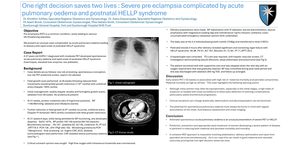
•At 31 weeks 6 days, while being admitted for BP monitoring, she developed dyspnea.   SaO2 >87%.  BP profile 145-156 systolic/94-102 diastolic.   Biochemistry workup:    Hb 107 , platelets 82, ALT 40, creatinine 70 ,PT 9.6 ,APTT19.4. PCR 138 , sFlt-1Plgf ratio 102.  Worsening proteinuria PCR 138mg/mmol.  Viral screening -ve. Urgent CXR, ECG, bedside echocardiogram were performed. CXR revealed severe pulmonary oedema (see Fig 1 ).
•Critical outreach opinion was sought.  High flow oxygen with intravenous frusemide was commenced. 
•Delivery preparations were made:  BP stabilization with IV labetalol; steroid administration; seizure prophylaxis with magnesium loading (4g) and maintenance (1g/hr) infusion; antibiotic cover; uncomplicated emergency caesarean section birth undertaken.  
•The baby was at the 4.4 individualized growth centile (1540g) and transferred to level 3 NICU.
•Postnatal bloods 4 hours after delivery revealed significant and worsening organ failure with HELLP syndrome: Hb 98, Plt 47, ALT 787, Bilirubin 41, Cr 80 , PT 11,APTT 28.5. 
•A hepatologist was consulted.   ITU care was required, with hepatic vascular screen, CT investigation demonstrating pleural effusions, basal atelectasis and pneumonia (see Fig 2).
•The patient recovered well with supportive care and was stepped down the next day with an oxygen requirement that was gradually weaned. BP was controlled during post natal period and she was discharged with labetalol 200 mg TDS and follow up arranged.

Discussion

Early severe PET (<32 weeks) is associated with high risk of  maternal mortality and perinatal compromise.   Maternal mortality as high as 20 fold.1  This case highlights the following learning points:

•Although many women may often be asymptomatic, especially in the initial stages, a high index of suspicion is needed with close surveillance to allow early detection of evolving complications, particularly subtle biochemical progression.

•Clinical situations can change drastically; deterioration and decompensation can be fulminant.

•The potential for spontaneous pulmonary oedema must always be borne in mind with regular auscultation of the chest, fluid balance assessment and chest imaging.

Conclusion

•Antenatal spontaneous acute pulmonary oedema is an unusual presentation of severe PET or HELLP.

•Prediction, prevention and appropriate early treatment of women developing a severe pattern of disease is pertinent in reducing both maternal and perinatal morbidity and mortality. 

•A cohesive MDT approach is imperative involving stabilization, delivery optimization and input from specialist services simultaneously.   As in our case this can result in good maternal and neonatal outcomes proving that one right decision saves two lives.


Copyright © 2015-2026, ePostersLive® SciGen is a registered trademark of SciGen Technologies S.A. Patent pending.

This website and third-party tools we use rely on cookies for the best user experience. By selecting "I agree", you agree to cookie usage as described in our Privacy Policy.