2,281 posters, 63 videos, 56 audios, 43 topics, 18 sessions, 6,174 authors, 2,323 institutions
ePostersLive by SciGen Technologies S.A. All rights reserved.
RCOG World Congress 2025
June 23 - 25, 2025 | ExCeL London, UK
EP.1651
Chylopericardium in pregnancy with cardiac tamponade and subsequent pre-eclampsia
Poster Presenter
Authors
Affiliations
Part of Topic
Maternal medicine
Chylopericardium in pregnancy with cardiac tamponade and subsequent pre-eclampsia
V Su1, S Rajendran1
1 Department of Obstetrics and Gynaecology, Royal Prince Alfred Hospital, Sydney, Australia
Objective:
To highlight the associated complications of chylopericardium in pregnancy
Case report:
A 39-year-old primigravid presents at 24+6 weeks gestation for routine antenatal care with new peripheral swelling and static fundal heights. On further history, she reported a 1-month history of intermittent chest pain, cough and orthopnoea. Past medical history included benign congenital pericardial cysts identified 2 years prior on routine chest x-ray performed for visa requirements, for which no further follow-up was recommended. Combined first trimester and preeclampsia risk screening were not completed.
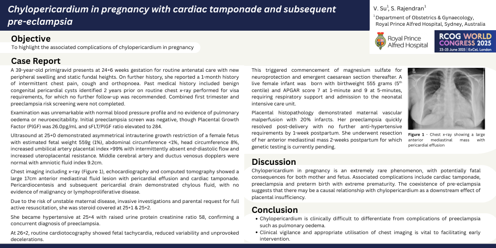
Examination was unremarkable with normal blood pressure profile and no evidence of pulmonary oedema or neuroexcitability. Initial preeclampsia screen was negative, though Placental Growth Factor (PlGF) was 26.0pg/mL and sFLT/PlGF ratio elevated to 284. Ultrasound at 25+0 demonstrated asymmetrical intrauterine growth restriction of a female fetus with estimated fetal weight 559g (1%), abdominal circumference <1%, head circumference 8%, increased umbilical artery placental index >99% with intermittently absent end-diastolic flow and increased uteroplacental resistance. Middle cerebral artery and ductus venosus dopplers were normal with amniotic fluid index 9.2cm. Chest imaging including x-ray, echocardiography and computed tomography showed a large 17cm anterior mediastinal fluid lesion with pericardial effusion and cardiac tamponade. Pericardiocentesis and subsequent pericardial drain demonstrated chylous fluid, with no evidence of malignancy or lymphoproliferative disease. Due to the risk of unstable maternal disease, invasive investigations and parental request for full active resuscitation, she was steroid covered at 25+1 & 25+2.
She became hypertensive at 25+4 with raised urine protein creatinine ratio 58, confirming a concurrent diagnosis of preeclampsia. At 26+2, routine cardiotocography showed fetal tachycardia, reduced variability and unprovoked decelerations. This triggered commencement of magnesium sulfate for neuroprotection and emergent caesarean section thereafter. A live female infant was born with birthweight 555 grams (5th centile) and APGAR score 7 at 1-minute and 9 at 5-minutes, requiring respiratory support and admission to the neonatal intensive care unit.
Placental histopathology demonstrated maternal vascular malperfusion with 20% infarcts. Her preeclampsia quickly resolved post-delivery with no further anti-hypertensive requirements by 1-week postpartum. She underwent resection of her anterior mediastinal mass 2-weeks postpartum for which genetic testing is currently pending.
Discussion:
Chylopericardium in pregnancy is an extremely rare phenomenon, with potentially fatal consequences for both mother and fetus. Associated complications include cardiac tamponade, preeclampsia and preterm birth with extreme prematurity. The coexistence of pre-eclampsia suggests that there may be a causal relationship with chylopericardium as a downstream effect of placental insufficiency.
Conclusions:
Chylopericardium is clinically difficult to differentiate from complications of preeclampsia such as pulmonary oedema. Clinical vigilance and appropriate utilisation of chest imaging is vital to facilitating early intervention.