1,830 posters, 67 videos, 75 audios, 29 topics, 22 sessions, 4,727 authors, 1,697 institutions
ePostersLive by SciGen Technologies S.A. All rights reserved.
RCOG World Congress 2023
June 12 - 14, 2023 | London, UK

EP.0726
A Case of a Missing Pregnancy of Unknown Location
Poster Presenter
Authors
Affiliations
Part of Topic
Early Pregnancy Complications and Gynaecology
A CASE OF PULMONARY CHORIOCARCINOMA MASKING AS A PREGNANCY OF UNKNOWN LOCATION
Objective
To discuss the investigation and management of a case of presumed pregnancy of unknown location (PUL).
To explore the diagnostic role of serial serum beta-hCG monitoring given negative non-invasive and invasive investigations.
To highlight the importance of working as a multi-disciplinary team (MDT) when faced with a complex diagnostic scenario.
Case Report
A 31-year-old lady with no significant past medical history, (one previous elective Caesarean section for breech and conservative management of early miscarriage, ex smoker), was referred to the EPAU for an ultrasound scan due to vaginal bleeding and pelvic pain. Her last menstrual period was approximately seven weeks prior. There was no evidence of an intrauterine pregnancy on imaging therefore serum beta-hCGs were arranged and revealed a rising trend with an initial beta-hCG of 19950 IU/L. Interval repeat ultrasound revealed no obvious pregnancy, intra-uterine or ectopic otherwise. A diagnostic laparoscopy and evacuation of the uterus was performed. No ectopic pregnancy was visualised during laparoscopy, however a small amount of material thought to be potential products of conception were evacuated from the uterus and sent for urgent histopathological examination.
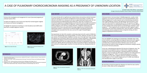
Post-operatively, her beta-hCGs revealed a continuing rising trend with a peak at 30194 IU/L. Prior to the histopathology results being available, a CT thorax, abdomen and pelvis (CT TAP) was ordered. This demonstrated a 10x8x6cm necrotic mass in the right lower lung, invading the adjacent liver and thoracic inferior vena cava with no distant metastases, in keeping with an image-based diagnosis of Primary Pulmonary Choriocarcinoma (PCC). Subsequent histopathology results from the surgical evacuation confirmed no trophoblastic tissue. The patient was urgently referred to Charing Cross where she started emergency chemotherapy and continues to undergo regular treatment. Her beta-hCG level has subsequently fallen to 8IU/L.
Discussion
Choriocarcinoma is rare occurring in 1:50,000 pregnancies, usually a molar pregnancy, occurring due to abnormality in the syncytiotrophoblastic cells 1. Although aggressive it usually responds well to chemotherapy and patients have a good prognostic outcome. Non-gestational choriocarcinoma is even rarer with only a handful of reported cases of PPC. The consensus of these reports are that there is no standardised treatment for PCC, it can be difficult to treat surgically with varying response to chemotherapy, resulting in a poor prognosis 2, 3. These diagnoses were considered only after negative surgical intervention and continually rising beta-hCGs. Given the patient’s response to chemotherapy, previous pregnancy history and genetic testing, she had a diagnosis of gestational choriocarcinoma.
Conclusion
This case highlights the importance of maintaining a ‘helicopter view’ of the clinical situation, considering differential diagnoses (particularly with negative imaging) and utilising multi-disciplinary discussions and case review. Often serial beta-hCGs are performed without definitive action and this case demonstrates the need for timely management and decision making to identify the source of hormone production.
In this case, undertaking post-operative hCGs became of diagnostic value. The continued rise prompted the notion to utilise further imaging to assess for extra-pelvic sources of hCG production, ultimately leading to the definitive diagnosis. Using risk models (eg. M4 model) is helpful to risk stratify patients to help guide management 4. There is no standardised guidance on further investigation of a PUL with a negative ultrasound, therefore consideration should be given to if non-invasive imaging such as CT or MRI, may initially be more beneficial over surgery in such cases.