1,830 posters, 67 videos, 75 audios, 29 topics, 22 sessions, 4,727 authors, 1,697 institutions
ePostersLive by SciGen Technologies S.A. All rights reserved.
RCOG World Congress 2023
June 12 - 14, 2023 | London, UK

EP.0422
Multiple chorioangioma syndrome - a rare form of placenta chorioangioma causing non-immune hydrops
Poster Presenter
Authors
Wai Lam Chan, Ying Tsz Viola Chan, Wai Kuen Yung, Lo Ka Chan, Wai Lam Lau, Chui San Zara Tsang, Mei Ching Chan, Hoi Wing Yuen, Wing Cheong Leung
Affiliations
Department of Obstetrics and Gynaecology, Kwong Wah Hospital, Department of Pathology, Kwong Wah Hospital, Department of Paediatrics and Adolescent Medicine, Kwong Wah Hospital
Part of Topic
Imaging in Obstetrics
Introduction
A case of hydrops fetalis caused by multiple chorioangioma syndrome presented at third trimester and resulted in a favourable neonatal outcome is reported. Sonographic appearance of the affected placenta and its role in antenatal diagnosis of the condition is discussed.
Case report
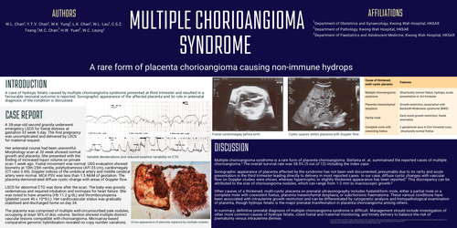
A 38-year-old second gravida underwent emergency LSCS for foetal distress at gestation 33 week 5 day. The first pregnancy was uncomplicated and delivered by LSCS for maternal request. Her antenatal course had been uneventful. Morphology scan at 20 week showed normal growth and placenta. She presented with the finding of increased liquor volume on private scan 1 week ago. Foetal movement was normal. USG evaluation showed biometry at 10th-25th centile, polyhydramnios (AFI 35 cm), cardiomegaly (CT ratio 0.69). Doppler indices of the umbilical artery and middle cerebral artery were normal. MCA PSV was less than 1.5 MoM of gestation. The placenta demonstrated diffuse cystic change with areas of Doppler flow.
LSCS for abnormal CTG was done after the scan. The baby was grossly oedematous and required intubation and inotropes for heart failure. She was noted to have anaemia (Hb 11.2 g/dL) and thrombocytopenia (platelet count 46 x 10^9/L). Her cardiovascular status was gradually stabilised and discharged home on day 34.
The placenta was comprised of multiple well-circumscribed pale nodules, occupying at least 50% of disc volume. Section showed multiple distinct vascular lesions compatible with chorioangioma. Microarray-based comparative genomic hybridisation revealed no copy number variations.
Discussion
Multiple chorioangioma syndrome is a rare form of placenta chorioangioma. Stefania et. al. summarised the reported cases of multiple chorioangioma.The overall survival rate was 38.5% (5 out of 13) including the index case. Sonographic appearance of placenta affected by the syndrome has not been well documented, presumably due to its rarity and acute presentation in the third trimester leading directly to delivery in most reported cases. In our case, diffuse cystic changes with vascular flow on Doppler studies were shown, whereas hypertrophic or slightly thickened appearance has been reported.This discrepancy can be attributed to the size of chorioangioma nodules, which can range from 1-2 mm to macroscopic growth.
Other causes of a thickened, multi-cystic placenta on prenatal ultrasonography includes hydatidiform mole, either a partial mole or a complete mole with coexistent foetus, placenta mesenchymal dysplasia, or subchorionic haematoma. These various conditions have been associated with intrauterine growth restriction and can be differentiated by cytogenetic analysis and histopathological examination of placenta, though hydrops fetalis is the major prenatal manifestation in placenta chorioangioma among others.
In summary, definitive prenatal diagnosis of multiple chorioangioma syndrome is difficult. Management should include investigation of other more common causes of hydrops fetalis, close foetal and maternal monitoring, and timely delivery to balance the risk of prematurity versus intrauterine demise.