1,830 posters, 67 videos, 75 audios, 29 topics, 22 sessions, 4,727 authors, 1,697 institutions
ePostersLive by SciGen Technologies S.A. All rights reserved.
RCOG World Congress 2023
June 12 - 14, 2023 | London, UK

EP.0418
Prenatal diagnosis of Cerebriform Sebaceous Naevus with Three-Dimensional (3D) ultrasound technology
Poster Presenter
Authors
Affiliations
Part of Topic
Imaging in Obstetrics
Prenatal diagnosis of Cerebriform Sebaceous Naevus with Three-dimensional (3D) ultrasound technology
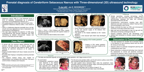
First described in 1895, sebaceous naevus (SN) is a congenital hamartoma of the pilosebaceous follicular unit. It is an uncommon type of birthmark that arises in about 0.3% of neonates. Cerebriform SN (CSN) is a rare morphological variant of SN, which was first reported in 1998. SN is usually reported to be present at birth however, it is more likely to be present antenatally. Due to the flat and smooth waxy surface presentation of SN in neonates, these lesions tend to be overlooked during prenatal ultrasound scans. CSN is usually not associated with extracutaneous manifestations, making it more challenging to detect on prenatal ultrasound scans.
We present a case of a multigravida who was referred to our Maternal Fetal Medicine (MFM) Service following for further evaluation of an abnormal ultrasound at 24 weeks of gestation which revealed abnormal bilateral fetal auricular masses. Serial obstetric ultrasound scans by our MFM specialists, with the aid of three-dimensional (3D) technology, were able to further characterise the lesion. The remainder of the fetal morphology was unremarkable. After delivery, the lesions on the neonate had the characteristic appearance of CSN at the locations visualised on the prenatal 3D ultrasound imaging. This diagnosis was subsequently confirmed to be CSN on biopsy.
Multiple specialities, including dermatology, plastic surgery, ear, nose and throat, genetics, cardiology and ophthalmology, assessed the baby and excluded associated syndromes with SN. Comprehensive examinations including brain MRI, abdominal and spine ultrasound, newborn hearing check and echocardiogram were normal. Baby was well and discharged on day 6 of age with the CSN lesions remaining unchanged at day 40. Ongoing outpatient follow up with dermatology and plastics were scheduled to consider surgical removal of the CSN.
The recent advances in 3D ultrasound technology have facilitated the increase in the detection rates of subtle fetal abnormalities, especially of the fetal face and profile. Whilst it is challenging to diagnose lesions such as SN prenatally, the utility of 3D ultrasound technology enabled the obstetric team to further characterise the lesion and rule out any associated syndromes or sinister causes associated with the lesions. It aided us in narrowing down the diagnosis to benign-causing lesions such as SN. Hence, the obstetric team were able to guide the mother in the management and outcome of the pregnancy.
To the best of our knowledge, this is the first case report illustrating the detection of CSN using 3D prenatal ultrasound, with CSN manifesting as bilateral infra-auricular masses and tumours in the facial and neck region. This case highlights the valuable role of prenatal imaging in the detection of rare cutaneous lesions with the aid of 3D ultrasound technology. Detection allows for postnatal management planning with multidisciplinary team input. Recent advances in ultrasound technology have improved improved the assessment of normal and abnormal structures, enhancing the detection rates of subtle fetal abnormalities, especially in the evaluation of the fetal face and profile.