1,830 posters, 67 videos, 75 audios, 29 topics, 22 sessions, 4,727 authors, 1,697 institutions
ePostersLive by SciGen Technologies S.A. All rights reserved.
RCOG World Congress 2023
June 12 - 14, 2023 | London, UK

EP.0368
Management of a vaginal stone in a patient with cerebral palsy
Poster Presenter
Authors
Rebecca Bennewith, Hafiza Saima Naseem, Radhika Padmagirison
Affiliations
Part of Topic
Urogynaecology/Pelvic Floor
Management of a vaginal stone in a patient with cerebral palsy
A Case Report
Introduction
Primary vaginal stones are rare diagnoses which are usually seen in women with underlying urogenital tract abnormalities when a stone develops because of urinary stasis in the vagina (Xu et al., 2020). Secondary vaginal calculi usually develop from the exposure of urine to retained foreign bodies within the vagina, such as synthetic mesh (Winkelman et al., 2016). Previously published case reports of vaginal stones in such women with prolonged immobility have predominantly been managed by minimally invasive, trans-vaginal stone removal (Aronson et al., 2020). This is the first known case of a patient requiring a hysterectomy in order to access the vaginal vault for removal of a large vaginal stone. Furthermore, this patient had the largest vaginal stone that has been reported in the literature
Case Presentation
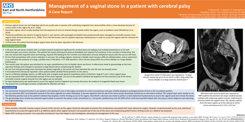
A 39-year-old care-home-resident with a complex medical background significant for cerebral palsy and epilepsy had multiple presentations to ED with abdominal pain and urinary retention. This patient had severe learning and physical disabilities and required full assistance in her activities of daily living. She was non-verbal with impaired vision and hearing, required PEG feeding for her nutrition and medication and was wheelchair-bound with the use of a full hoist. On her third presentation with urinary retention in one year, the urology registrar observed a bladder stone during a difficult catheterisation, and an abdominal x-ray confirmed the presence of a large, calcified mass in the pelvis. A CT KUB reported a 10cm calculus lying within the urinary bladder or a large bladder diverticulum. Two months later the patient was admitted for an open cystolithotomy, but no bladder stone was found. A referral was made to gynaecology as the intra-operative findings were thought to represent a large fibroid uterus compressing the bladder. No one from the gynaecology team examined the patient vaginally as she would not have tolerated this and she was not sexually active. A transabdominal ultrasound reported a 10cm pedunculated calcified fibroid posterior to the uterus. Due to conflicting radiology reports, an MRI pelvis was arranged under general anaesthesia which confirmed a large 8.5 x 8.5 x 12cm vaginal stone. Our pre-operative MDT recommended removal of the stone vaginally but due to the patient’s bilateral hip dysplasia and the enormous size of the stone, minimally invasive surgical removal from the vagina was not possible. An open hysterectomy was performed to access the vaginal vault to fragment and remove the stone. No evidence of urogenital fistula was found. The patient had a prolonged recovery complicated by infection and ileus.
Discussion
The mechanism of stone formation in our patient is the pooling of urine in the vagina secondary to urinary incontinence and poor mobility leading to prolonged periods of time in the recumbent position. Our pre-operative MDT recommended removal of the stone vaginally but when attempted, it became apparent that the size of the stone would necessitate removal by an alternative method. The surgical team were unable to use the lithotomy position due to the patient’s bilateral hip dysplasia, further limiting access to the vagina. Henceforth a decision was made to perform a total abdominal hysterectomy to access the vaginal vault and remove the stone. The gynaecology team missed two potential opportunities to examine the patient under anaesthesia (i.e. during the cystolithotomy and the MRI) which could have possible helped with securing an earlier diagnosis and improving the surgical planning.
Conclusion
When possible, minimally invasive surgical removal of the calculus via the vagina should be attempted to prevent the complications associated with major abdominal surgery. However, as demonstrated by this case, abdominal hysterectomy to access the vaginal vault is an effective option when vaginal removal is not possible due to the size of the stone and complicating patient factors limiting access (i.e. bilateral hip dysplasia). Better MDT working and earlier cross-speciality involvement may have helped in the investigation, planning and management of this case.