1,830 posters, 67 videos, 75 audios, 29 topics, 22 sessions, 4,727 authors, 1,697 institutions
ePostersLive by SciGen Technologies S.A. All rights reserved.
RCOG World Congress 2023
June 12 - 14, 2023 | London, UK

EP.1369
Antenatal diagnosis and neonatal outcome of Situs Solitus, Dextrocardia: A case report
Part of Topic
Imaging in Obstetrics
ANTENATAL DIAGNOSIS AND NEONATAL OUTCOME OF SITUS SOLITUS, DEXTROCARDIA
A CASE REPORT
J.S.Manamperi1
1 Intern medical officer, Teaching Hospital Peradeniya
INTRODUCTION
Dextrocardia is a rare congenital abnormality in the general population with an estimated incidence of 1 in 12,000 live births. Isolated dextrocardia, the condition of right-sided heart with situs solitus most commonly referred to as dextroversion
CASE REPORT
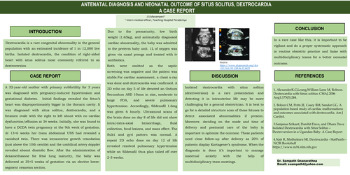
A 32-year-old mother with primary subfertility for 3 years was diagnosed with pregnancy-induced hypertension and gestational diabetes. Initial findings revealed the fetus’s heart was disproportionately bigger in the thoracic cavity. It was diagnosed with situs solitus, dextrocardia, and a foramen ovale with the right to left shunt with no cardiac dysfunction/effusion at 34 weeks. Initially, she was found to have a DCDA twin pregnancy at the 9th week of gestation. At 13+6 weeks her trans abdominal USS had revealed a vanished twin. There was intrauterine growth retardation (just above the 10th centile) and the umbilical artery doppler revealed absent diastolic flow. After the administration of dexamethasone for fetal lung maturity, the baby was delivered at 35+5 weeks of gestation via an elective lower-segment cesarean section.
Due to the prematurity, low birth weight (1.65kg), and antenatally diagnosed cardiac abnormality, the baby was admitted to the preterm baby unit. 1L of oxygen was given via nasal prongs and treated with iv antibiotics.
Both were omitted as the septic screening was negative and the patient was stable.For cardiac assessment, a chest x-ray was done and dextrocardia was confirmed. A 2D echo on day 5 of life denoted an Ostium Secundum ASD 10mm in size, moderate to large PDA, and severe pulmonary hypertension. Accordingly, Sildenafil 1.6mg was given 6 hourly. Ultrasound scans of the brain done on day 8 of life did not show intra/extra-axial hemorrhage, fluid collection, focal lesions, and mass effect. The Sulci and gyri pattern was normal. A repeat 2D echo done on day 12 of life revealed resolved pulmonary hypertension while on Sildenafil thus plan tailed off over 2-3 weeks.
DISCUSSION
Isolated dextrocardia with situs solitus (dextroversion) is a rare presentation and detecting it in intrauterine life may be more challenging for a general obstetrician. It is best to go for a detailed structure scan of these fetuses to detect associated abnormalities if present. Moreover, deciding on the mode and time of delivery and postnatal care of the baby is important to optimize the outcome. These patients need close follow-up after delivery as 20% of patients display Kartagener’s syndrome. When the diagnosis is done it’s important to manage maternal anxiety with the help of multidisciplinary team meetings.
CONCLUSION
In a rare case like this, it is important to be vigilant and do a proper systematic approach in routine obstetric practice and liaise with multidisciplinary teams for a better neonatal outcome.
REFERENCES
1. AlexanderK.C,Leung,William Lane M, Robson. Dextrocardia with Sinus solitus: CMAJ.2006 Aug1;175(3):244.
2. Bohun CM, Potts JE, Casey BM, Sandor GG. A population-based study of cardiac malformations and outcomes associated with dextrocardia. Am J Cardiol.
3.Sanjanaa Srikant, Darshit Dave, and Dhara Dave
Isolated Dextrocardia with Situs Solitus – Dextroversion in a Ugandan Baby: A Case Report
4.Nair R, Muthukuru SR. Dextrocardia - StatPearls - NCBI Bookshelf
https://www.ncbi.nlm.nih.gov