1,830 posters, 67 videos, 75 audios, 29 topics, 22 sessions, 4,727 authors, 1,697 institutions
ePostersLive by SciGen Technologies S.A. All rights reserved.
RCOG World Congress 2023
June 12 - 14, 2023 | London, UK

EP.1126
Newborn with a congenital ovarian cyst – A rare clinical entity
Poster Presenter
Part of Topic
Early Pregnancy Complications and Gynaecology
Newborn with a Congenital Ovarian Cyst – A rare clinical entity
V.J. Meegoda1, K.G. Hewawitharana1
1University professorial unit of Obstetrics & Gynecology, Colombo South Teaching Hospital, Colombo, Sri Lanka.
INTRODUCTION
§Congenital ovarian cysts are rare, with less than a hundred reported cases.
§These can be diagnosed through antenatal ultrasonography (USS).
§Majority are benign, functional cysts of graafian origin and have an excellent prognosis.
§However, complications such as torsion, rupture, hemorrhage, urinary tract obstruction, thoracic compression with pulmonary hypoplasia, and even sudden death are possibilities, especially in large cysts which may occupy a majority of abdominal cavity.
§All complicated ovarian cysts and simple cysts >5cm should be intervened.
CASE PRESENTATION
•Here we report a case of an infant with a unilateral, progressively enlarging ovarian cyst diagnosed during the third trimester by antenatal USS.
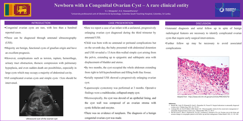
•Child was born with no antenatal or perinatal complications but on the seventh day, the baby presented with abdominal distention and USS revealed a 13.6cm thin-walled simple cyst arising from the pelvis, extending up to epigastric and subhepatic area with displacement of bladder and uterus.
•By two months, the cyst occupied the whole abdomen extending from right to left hypochondrium and filling both iliac fossae.
•Serially repeated USS showed a progressively enlarging ovarian cyst.
•Laparoscopic cystectomy was performed at 3 months. Operative findings were a multilocular, collapsed empty cyst.
•Microscopically, the cyst was devoid of an epithelial lining, and the cyst wall was composed of an ovarian stroma with cystic follicles and oocytes.
•There was no evidence of neoplasm. The diagnosis of a benign congenital ovarian cyst was made.
DISCUSSION
•Antenatal diagnosis and serial follow up in spite of benign radiological features are necessary to identify complicated ovarian cysts that require early surgical interventions.
•Further follow up may be necessary to avoid associated complications