296 posters, 7 videos, 13 audios, 14 topics, 10 sessions, 1,019 authors, 259 institutions
ePostersLive by SciGen Technologies S.A. All rights reserved.
RCOphth Annual Congress 2026
18 - 21 May, 2026 | Manchester Central, Manchester

P240
Optic Neuropathy Due to Idiopathic Spontaneous Gyrus Rectus Herniation in an Adolescent: Clinical and Imaging Insights
Poster Presenter
Authors
Part of Topic
Neuro ophthalmology
Background:
Posterior to the gyrus rectus lies the intracranial optic nerve and anterior chiasm,
and compression is usually neoplastic [1]
• Spontaneous herniation of the gyrus rectus is uncommon [2] and may present
with visual symptoms.
• We report a rare case of spontaneous idiopathic gyrus rectus herniation in
an adolescent, resulting in hemi-chiasmal compression
Case presentation:
17-year-old female with a one-day history of acute right visual loss
• Associated mild retro-orbital discomfort
• Previously healthy; no relevant medical or family history
• Visual acuity: Right eye (RE) LogMAR 1.24 (1.2 pinhole), Left eye (LE) 0.2
• Colour vision: RE 2/17, LE 17/17 (Ishihara)
• No relative afferent pupillary defect (RAPD); full extraocular movements
• Fundoscopy and optical coherence tomography (OCT) of disc and macula were
normal
Investigations and imaging:
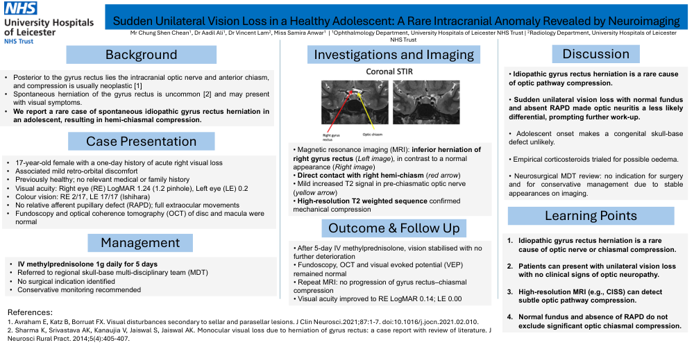
Magnetic resonance imaging (MRI): inferior herniation of
right gyrus rectus (Left image), in contrast to a normal
appearance (Right image)
• Direct contact with right hemi-chiasm (red arrow)
• Mild increased T2 signal in pre-chiasmatic optic nerve
(yellow arrow)
• High-resolution T2 weighted sequence confirmed
mechanical compression
Management:
IV methylprednisolone 1g daily for 5 days
• Referred to regional skull-base multi-disciplinary team (MDT)
• No surgical indication identified
• Conservative monitoring recommended
Outcome & Follow-up:
After 5-day IV methylprednisolone, vision stabilised with no
further deterioration
• Fundoscopy, OCT and visual evoked potential (VEP)
remained normal
• Repeat MRI: no progression of gyrus rectus–chiasmal
compression
• Visual acuity improved to RE LogMAR 0.14; LE 0.00
Discussion:
Idiopathic gyrus rectus herniation is a rare cause
of optic pathway compression.
• Sudden unilateral vision loss with normal fundus
and absent RAPD made optic neuritis a less likely
differential, prompting further work-up.
• Adolescent onset makes a congenital skull-base
defect unlikely.
• Empirical corticosteroids trialed for possible oedema.
• Neurosurgical MDT review: no indication for surgery
and for conservative management due to stable
appearances on imaging.
Learning points:
1. Idiopathic gyrus rectus herniation is a rare
cause of optic nerve or chiasmal compression.
2. Patients can present with unilateral vision loss
with no clinical signs of optic neuropathy.
3. High-resolution MRI (e.g., CISS) can detect
subtle optic pathway compression.
4. Normal fundus and absence of RAPD do not
exclude significant optic chiasmal compression