296 posters, 7 videos, 13 audios, 14 topics, 10 sessions, 1,019 authors, 259 institutions
ePostersLive by SciGen Technologies S.A. All rights reserved.
RCOphth Annual Congress 2026
18 - 21 May, 2026 | Manchester Central, Manchester

P236
Association of retinal and OCT age gap and multiple sclerosis
Poster Presenter
Authors
Part of Topic
Neuro ophthalmology
Backgroud
Multiple sclerosis (MS) is a chronic autoimmune disease characterised by demyelination and progressive neurodegeneration of the central nervous system.
•Epidemiological trends show rising prevalence of MS among older adults [1]. However, chronological age fails to capture the variability in biological ageing among individuals.
•Existing biomarkers of biological ageing in MS such as telomere length [2] cellular senescence, and MRI-derived brain age are invasive, costly, and impractical at scale.
•The retina offers a non-invasive window to the CNS via fundus photography and OCT imaging.
•Retinal age gap (the difference between AI-predicted retinal age and chronological age) is an emerging biomarker of biological ageing.
•This study investigates whether retinal and OCT age gaps are elevated in individuals with MS compared to matched controls.
Methods
•This study used data from the UK Biobank, a prospective population-based cohort of over 500,000 recruited between 2006 and 2010.
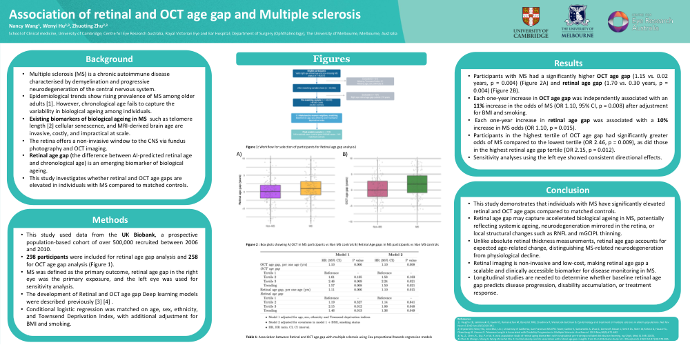
•After applying inclusion criteria, 298 participants were included in retinal age gap analysis and 258 for OCT age gap analysis (Figure 1).
•MS was defined as the primary outcome, with retinal age gap in the right eye as the primary exposure; the left eye was used for sensitivity analysis.
•Retinal and OCT age gaps were calculated using previously described deep learning models [3,4].
•Conditional logistic regression was matched on age, sex, ethnicity, and Townsend Deprivation Index, with additional adjustment for BMI and smoking.
Results
•Participants with MS had a significantly higher OCT age gap (1.15 vs. 0.02 years, p = 0.004) (Figure 2A) and retinal age gap (1.70 vs. 0.30 years, p = 0.004) (Figure 2B).
•Each one-year increase in OCT age gap was independently associated with an 10%increase in the odds of MS (OR 1.10, 95% CI, p = 0.008) after adjustment for BMI and smoking.
• Each one-year increase in retinal age gap was associated with a 10% increase in MS odds (OR 1.10, p = 0.015).
•Participants in the highest tertile of OCT age gap had significantly greater odds of MS compared to the lowest tertile (OR 2.46, p = 0.009), as did those in the highest retinal age gap tertile (OR 2.15, p = 0.012).
•Sensitivity analyses using the left eye showed consistent directional effects.
Conclusion
•This study demonstrates that individuals with MS have significantly elevated retinal and OCT age gaps compared to matched controls.
•Retinal age gap may capture acceleratedbiological ageing in MS, potentially reflecting systemic ageing, neurodegeneration mirrored in the retina, or local structural changes such as RNFL and mGCIPL thinning.
•Unlike absolute retinal thickness measurements, retinal age gap accounts for expected age-related change, distinguishingMS-related neurodegeneration from physiological decline.
•Retinal imaging is non-invasive and low-cost, making retinal age gap a scalable and clinically accessible biomarker for disease monitoring in MS.
•Longitudinal studies are needed to determine whether baseline retinal age gap predicts disease progression, disability accumulation, or treatment response.