215 posters, 5 topics, 22 sessions, 596 authors, 283 institutions
ePostersLive by SciGen Technologies S.A. All rights reserved.
Obstetric Anaesthesia Annual Scientific Meeting 2025
May 15 - 16, 2025 | ICC, 2 Lanyon Place, Belfast BT1 3WH

6780188
Mechanical thrombectomy following stroke in second stage of labour
Poster Presenter
Part of Topic
Case report
Mechanical Thrombectomy Following Stroke in Second Stage of Labour
Dr Kathryn Shields ST4 Anaesthesia Resident, Dr Rina James Consultant Anaesthetist
Royal Jubilee Maternity Hospital, Belfast
Case Summary
31 year old female, para 0, 38+4 gestation with spontaneous
rupture of membranes. Past medical history of congenital
atrioventricular block, managed with transvenous pacemaker.
Uneventful antenatal course. Pre-pregnancy transthoracic
echocardiogram - low normal left ventricular systolic function
with no significant valve abnormalities. Pacemaker check at
approximately 26 weeks gestation: atrial tachycardia with 2:1 AV
block.
As first stage of labour progressed, analgesia was provided via
epidural with T6 bilateral sensory block. “Vagueness” reported 45
minutes into second stage. The patient was aphasic, with no
motor function in right arm, not following commands.
Observations were stable with the exception of tachycardia.
Epidural pump stopped immediately, cardiology and neurology
advice sought and emergency stroke team activated at 14:00.
Live infant delivered by forceps. Blood loss 1480ml due to atonic
uterus and trauma.
Stroke team assessed patient - GCS 11/15, partial gaze palsy,
minor facial palsy, aphasic, no motor function right arm - NIHSS
score 34 (severe stroke).
Management and Outcome
The patient was transferred via blue light ambulance to the main
hospital for imaging. CT brain - no haemorrhage, no established
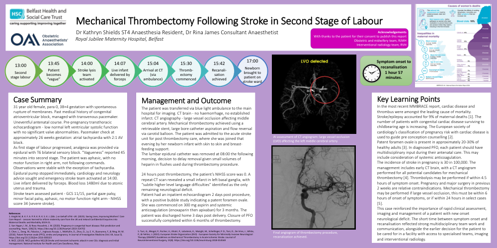
infarct. CT angiography - large vessel occlusion affecting middle
cerebral artery. Mechanical thrombectomy achieved using a
retrievable stent, large bore catheter aspiration and flow reversal
via carotid balloon. The patient was admitted to the acute stroke
unit for post thrombectomy care, where she was joined that
evening by her newborn infant with skin to skin and breast-
feeding support.
The lumbar epidural catheter was removed at 08:00 the following
morning, decision to delay removal given small volumes of
heparin in flushes used during thrombectomy procedure.
24 hours post thrombectomy, the patient’s NIHSS score was 0. A
repeat CT scan revealed a small infarct in left basal ganglia, with
“subtle higher level language difficulties” identified as the only
remaining neurological deficit.
Patient had an inpatient echocardiogram 2 days post procedure,
with a positive bubble study indicating a patent foramen ovale.
She was commenced on 300 mg aspirin and systemic
anticoagulation (enoxaparin then apixaban) for 3 months. The
patient was discharged home 3 days post delivery. Closure of PFO
successfully completed within 6 months of thrombectomy.
Key Learning Points
In the most recent MMBRACE report, cardiac disease and
thrombus were amongst the leading cause of mortality.
Stroke/epilepsy accounted for 9% of maternal deaths [1]. The
number of patients with congenital cardiac disease surviving to
childbearing age is increasing. The European society of
cardiology’s classification of pregnancy risk with cardiac disease is
used to guide pre conception counselling [2].
Patent foramen ovale is present in approximately 20-30% of
healthy adults [3]. In diagnosed PFO, each patient should have
multidisciplinary input during their antenatal care. This may
include consideration of systemic anticoagulation.
The incidence of stroke in pregnancy is 30 in 100,000. The
management includes early CT brain, with a CT angiogram
performed for all potential candidates for mechanical
thrombectomy [4]. Thrombolysis may be performed if within 4.5
hours of symptom onset. Pregnancy and major surgery in previous
2 weeks are relative contraindications. Mechanical thrombectomy
may be performed if large vessel occlusion, this must be within 6
hours of onset of symptoms, or if within 24 hours in select cases
[5].
This case reinforced the importance of rapid clinical assessment,
imaging and management of a patient with new onset
neurological deficit. The short time between symptom onset and
recanalisation reflected strong multidisciplinary team work and
communication, alongside the earlier decision for the patient to
be cared for in a facility with access to specialised teams, imaging
and interventional radiology.