215 posters, 5 topics, 22 sessions, 596 authors, 283 institutions
ePostersLive by SciGen Technologies S.A. All rights reserved.
Obstetric Anaesthesia Annual Scientific Meeting 2025
May 15 - 16, 2025 | ICC, 2 Lanyon Place, Belfast BT1 3WH

6780142
Rebound intracranial hypertension with papilledema following epidural blood patch for post-dural puncture headache in a postpartum patient
Poster Presenter
Part of Topic
Case report
Rebound Intracranial Hypertension with Papilloedema Following Epidural Blood Patch for Post-Dural Puncture Headache: A Case Report
Lakshmi Sagar Vijayakumar1, Nitesh Bargaje1, Preeti Nemade1 1Bedfordshire Hospitals NHS Foundation Trust
Introduction
Post-dural puncture headache (PDPH), a prevalent complication of neuraxial anaesthesia, particularly after an inadvertent dural puncture with an epidural needle, leads to intracranial hypotension, which is typically managed conservatively or with an epidural blood patch (EBP). While EBPs demonstrate efficacy, rare complications such as rebound intracranial hypertension (RIH) can manifest, causing persistent headaches and visual disturbances. RIH post-EBP can closely resemble PDPH, posing challenges in diagnosis and treatment [1]. We present a case of RIH accompanied by papilloedema following an EBP in a postpartum patient, emphasising the significance of early recognition and management.
Case Details
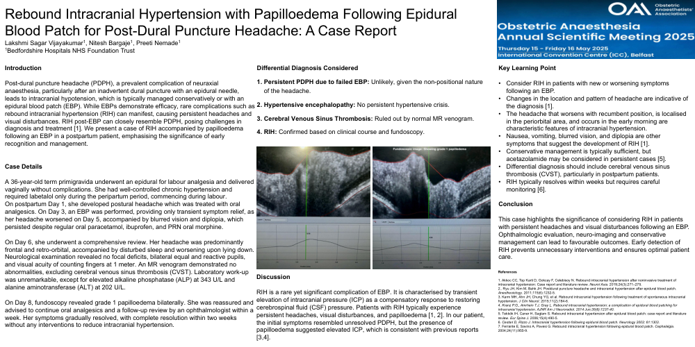
A 36-year-old term primigravida underwent an epidural for labour analgesia and delivered vaginally without complications. She had well-controlled chronic hypertension and required labetalol only during the peripartum period, commencing during labour. On postpartum Day 1, she developed postural headache which was treated with oral analgesics. On Day 3, an EBP was performed, providing only transient symptom relief, as her headache worsened on Day 5, accompanied by blurred vision and diplopia, which persisted despite regular oral paracetamol, ibuprofen, and PRN oral morphine. On Day 6, she underwent a comprehensive review. Her headache was predominantly frontal and retro-orbital, accompanied by disturbed sleep and worsening upon lying down. Neurological examination revealed no focal deficits, bilateral equal and reactive pupils, and visual acuity of counting fingers at 1 meter. An MR venogram demonstrated no abnormalities, excluding cerebral venous sinus thrombosis (CVST). Laboratory work-up was unremarkable, except for elevated alkaline phosphatase (ALP) at 343 U/L and alanine aminotransferase (ALT) at 202 U/L. On Day 8, fundoscopy revealed grade 1 papilloedema bilaterally. She was reassured and advised to continue oral analgesics and a follow-up review by an ophthalmologist within a week. Her symptoms gradually resolved, with complete resolution within two weeks without any interventions to reduce intracranial hypertension.
Differential Diagnosis Considered 1. Persistent PDPH due to failed EBP: Unlikely, given the non-positional nature of the headache. 2. Hypertensive encephalopathy: No persistent hypertensive crisis. 3. Cerebral Venous Sinus Thrombosis: Ruled out by normal MR venogram. 4. RIH: Confirmed based on clinical course and fundoscopy.
Discussion
RIH is a rare yet significant complication of EBP. It is characterised by transient elevation of intracranial pressure (ICP) as a compensatory response to restoring cerebrospinal fluid (CSF) pressure. Patients with RIH typically experience persistent headaches, visual disturbances, and papilloedema [1, 2]. In our patient, the initial symptoms resembled unresolved PDPH, but the presence of papilloedema suggested elevated ICP, which is consistent with previous reports [3,4].
Key Learning Point • Consider RIH in patients with new or worsening symptoms following an EBP. • Changes in the location and pattern of headache are indicative of the diagnosis [1]. • The headache that worsens with recumbent position, is localised in the periorbital area, and occurs in the early morning are characteristic features of intracranial hypertension. • Nausea, vomiting, blurred vision, and diplopia are other symptoms that suggest the development of RIH [1]. • Conservative management is typically sufficient, but acetazolamide may be considered in persistent cases [5]. • Differential diagnosis should include cerebral venous sinus thrombosis (CVST), particularly in postpartum patients. • RIH typically resolves within weeks but requires careful monitoring [6].
Conclusion
This case highlights the significance of considering RIH in patients with persistent headaches and visual disturbances following an EBP. Ophthalmologic evaluation, neuro-imaging and conservative management can lead to favourable outcomes. Early detection of RIH prevents unnecessary interventions and ensures optimal patient care.
References 1. Akkoc CC, Top Karti D, Gokcay F, Celebisoy N. Rebound intracranial hypertension after noninvasive treatment of intracranial hypotension: Case report and literature review. Neurol Asia. 2019;24(3):271–275. 2.. Ryu JH, Kim M, Bahk JH. Postdural puncture headache and intracranial hypertension after epidural blood patch. Anesthesiology. 2011;115(6):1232-5. 3. Karm MH, Ahn JH, Chung YG, et al. Rebound intracranial hypertension following treatment of spontaneous intracranial hypotension. J Clin Neurol. 2015;11(2):184-6. 4. Kranz P.G., Amrhein T.J, Gray L. Rebound intracranial hypertension: a complication of epidural blood patching for intracranial hypotension. AJNR Am J Neuroradiol. 2014 Jun;35(6):1237-40. 5. Tekkök IH, Caner H, Saglam S. Rebound intracranial hypertension after epidural blood patch: case report and literature review. Eur Spine J. 2006;15(4):490-5. 6. Cestari D, Rizzo J. Intracranial hypertension following epidural blood patch. Neurology 2003; 61:1303. 7. Ferrante E, Savino A, Pavesi G. Rebound intracranial hypertension following epidural blood patch. Cephalalgia. 2004;24(11):955-9.