293 posters, 56 videos, 24 topics, 13 sessions, 658 authors, 215 institutions
ePostersLive by SciGen Technologies S.A. All rights reserved.
ISMICS 2025 Annual Meeting
28 - 31 May, 2025 | Istanbul, Turkey

D143
The Role Of End-state Imaging In Enhancing Transcatheter Aortic Valve Implantation Outcomes And Procedural Success
Poster Presenter
Authors
Michael Wilson, Henry Wilson, Veronica Preda, Steven Agiasotis, Abirami Puvanendran, Terry Carney, Martin Ng
Affiliations
Surgical XR, 2 Technology Place, Macquarie Park, Sydney Australia., Faculty of Medicine, Health and Human Sciences, Macquarie University Macquarie Park, Sydney Australia and Macquarie University Hospital, 2 Technology Place, Macquarie Park, Sydney Australia, Faculty of Medicine, Health and Human Sciences, Macquarie University Macquarie Park, Sydney Australia, Surgical XR, 2 Technology Place, Macquarie Park. Faculty of Medicine, Health and Human Sciences, Macquarie University Macquarie Park, 2 Technology Place, Macquarie Park
Part of Topic
Education, Imaging and Facilitating Technologies
Background
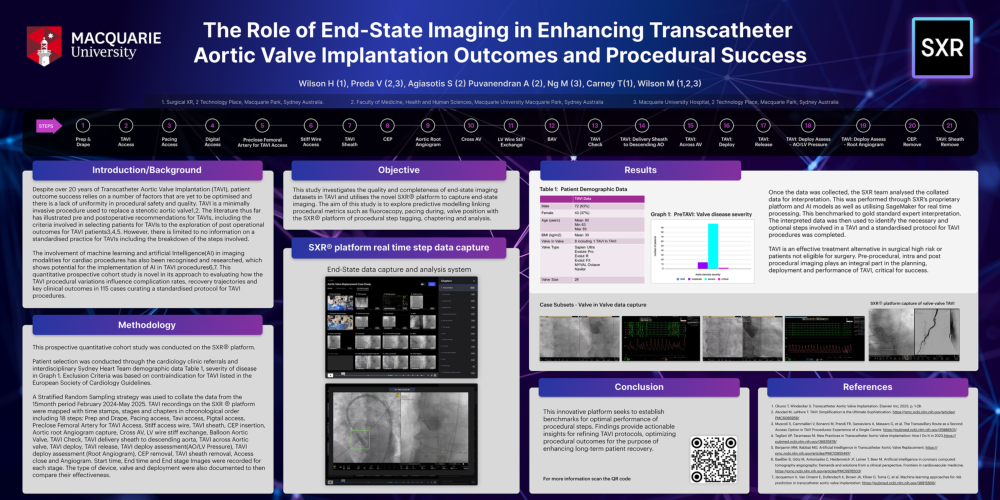
Despite over 20 years of Transcatheter Aortic Valve Implantation (TAVI), patient outcome success relies on a number of factors that are yet to be optimised and there is a lack of uniformity in procedural safety and quality. TAVI is a minimally invasive procedure used to replace a stenotic aortic valve1,2. The literature thus far has illustrated pre and postoperative recommendations for TAVIs, including the criteria involved in selecting patients for TAVIs to the exploration of post operational outcomes for TAVI patients3,4,5. However, there is limited to no information on a standardised practice for TAVIs including the breakdown of the steps involved. The involvement of machine learning and artificial Intelligence(AI) in imaging modalities for cardiac procedures has also been recognised and researched, which shows potential for the implementation of AI in TAVI procedures6,7. This quantitative prospective cohort study is novel in its approach to evaluating how the TAVI procedural variations influence complication rates, recovery trajectories and key clinical outcomes in >100 cases curating a standardised protocol for TAVI procedures.
Objective
This study investigates the quality and completeness of end-state imaging datasets in TAVI and utilises the novel SXR® platform to capture end-state imaging. The aim of this study is to explore predictive modelling linking procedural metrics such as fluoroscopy, pacing during, valve position with the SXR® platform of procedural step tagging, chaptering and analysis.
Methodology
This prospective quantitative cohort study was conducted on the SXR ® platform.
Patient selection was conducted through the cardiology clinic referrals and interdisciplinary Sydney Heart Team.
A Stratified Random Sampling strategy was used to collate the data from the 15month period February 2024-May 2025. TAVI recordings on the SXR ® platform were mapped with time stamps, stages and chapters in chronological order including 20 steps: Prep and Drape, Pacing access, Tavi access, Pigtail access, Preclose Femoral Artery for TAVI Access, Stiff access wire, TAVI sheath, CEP insertion, Aortic root Angiogram capture, Cross AV, LV wire stiff exchange, Balloon Aortic Valve, TAVI Check, TAVI delivery sheath to descending aorta, TAVI across Aortic valve, TAVI release, TAVI deploy assessment (AO/LV Pressure) and TAVI deploy assessment (Root Angiogram), CEP removal, TAVI sheath removal, Access close and Angiogram. Start time, End time and End stage Images were recorded for each stage. The type of device, valve and deployment were also documented to then compare their effectiveness.
Results: SXR® platform real time step data capture
Figure 1: TAVI chapters SXR layout
Figure 2: Timestamped ESI of valve deployment
Discussion/Conclusion
Structured ESI allows for clearer documentation and reproducibility across TAVI procedures. By identifying imaging targets at key steps, particularly valve deployment and post-op haemodynamics. The Platform provides a framework for retrospective quality assurance and training. These datasets can also support machine learning models that identify errors or predict outcomes.
SXR seeks to establish benchmarks for optimal performance of procedural steps. Findings provide actionable insights for refining TAVI protocols, optimizing procedural outcomes for the purpose of enhancing long-term patient recovery.
By embedding image capture into chaptered procedural timelines, this methodology enables better training, auditing, and future real-time support in structural heart intervention
Scan for more Information
Aortic Root Angio
Establish co-planar or 2-cusp view for valve orientation
Pigtail Access
Show pigtail in non- coronary sinus; used for valve alignment
CEP Filter Deployment
Show filter position in BCA and LCC
TAVI Delivery
Show valve mounted on delivery system in descending thoracic aorta
Valve Deployment
Capture deployment with pacing, angiographic positioning, hemodynamics
Post-Deployment Assessment
Confirm valve expansion, ARI, and assess for PVL