293 posters, 56 videos, 24 topics, 13 sessions, 658 authors, 215 institutions
ePostersLive by SciGen Technologies S.A. All rights reserved.
ISMICS 2025 Annual Meeting
28 - 31 May, 2025 | Istanbul, Turkey

D72
Cardiac Herniation Through The Chest Wall After Minimally Invasive Coronary Artery Bypass Surgery
Poster Presenter
Authors
Anıl Karaağaç, Gökhan Arslanhan, Murat Baştopcu, Hatice Doğusoy, Zeynep Sıla Özcan, Halim Ulugöl, Şahin Şenay, Cem Alhan, Ilgaz Doğusoy
Affiliations
Acıbadem Altunizade Hospital, Department of Anesthesiology, Istanbul, Turkey, Acıbadem Maslak Hospital, Department of Cardiovascular Surgery, Istanbul, Turkey, Acıbadem University School of Medicine, Department of Cardiovascular Surgery, Istanbul, Turkey, Acibadem Altunizade Hospital, Department of Thoracic Surgery, Istanbul, Turkey
Part of Topic
Coronary Revascularization
Video
Cardiac Herniation Through the Chest Wall After Minimally Invasive Coronary Artery Bypass Surgery
Background: Minimally invasive cardiac surgery is performed frequently in common practice, leading to an increased incidence of spesific complications related to the operative techniques. Cardiac herniation is a potential example of these. In this video, we present a patient with herniation of the cardiac apex through the chest wall after minimally invasive coronary artery bypass graft surgery.
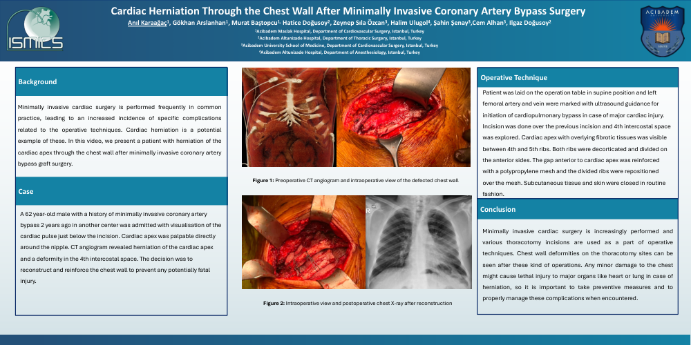
Methods: A 62 year-old male with a history of minimally invasive coronary artery bypass 2 years ago in another center was admitted with visualisation of the cardiac pulse just below the incision. Cardiac apex was palpable directly around the nipple. CT angiogram revealed herniation of the cardiac apex and a deformity in the 4th intercostal space. The decision was to reconstruct and reinforce the chest wall to prevent any potentially fatal injury.
Operative technique: The patient was laid on the operation table in supine position and left femoral artery and vein were marked with ultrasound guidance for initiation of cardiopulmonary bypass in case of major cardiac injury. Incision was done over the previous incision and 4th intercostal space was explored. Cardiac apex with overlying fibrotic tissues was visible between 4th and 5th ribs. Both ribs were decorticated and divided on the anterior sides. The gap anterior to cardiac apex was reinforced with a polypropylene mesh and the divided ribs were repositioned over the mesh. Subcutaneous tissue and skin were closed in routine fashion.
Conclusion: Minimally invasive cardiac surgery is increasingly performed and various thoracotomy incisions are used as a part of operative techniques. Chest wall deformities on the thoracotomy sites can be seen after these kind of operations. Any minor damage to the chest might cause lethal injury to major organs like heart or lung in case of herniation, so it is important to take preventive measures and to properly manage these complications when encountered.