136 posters, 2 topics, 27 sessions, 339 authors, 88 institutions
ePostersLive by SciGen Technologies S.A. All rights reserved.
2024 ACP Annual Session
NOVEMBER 6-9, 2024 | MARCO ISLAND, FL JW MARRIOT

13
Economical approach to obtaining full-arch implant scan with smartphone and deep learning: An in vitro investigation on trueness and precision
Poster Presenter
Authors
Fatma Mohamed, Zhaozhao Chen, Fei Liu , Berna Saglik , Gusatvo Mendonca, Junying Li
Affiliations
Part of Topic
Original Research
Economical approach to obtaining full-arch implant scan with smartphone and deep learning: An in vitro investigation on trueness and precision
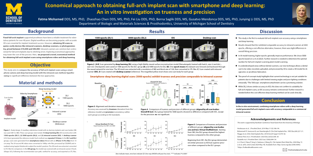
Fixed full-arch implant-supported prostheses have been a reliable treatment for edentulous patients for over 40 years. Digital workflows are now standard, with accurate 3D scans essential for implant treatment success. However, obtaining 3D scans requires costly devices like intraoral scanners, desktop scanners, or photogrammetry, priced between $10,000 and $40,000. Intraoral scanners are common but controversial for full-arch implants due to stitching errors. Exploring economical approaches may lower the cost of this process. Here, we proposed a novel, economical method for obtaining full-arch implant scans using smartphone video and deep learning.
This study aims to compare the accuracy of full-arch implant scans using a smartphone camera and deep learning model with the intraoral scan method, hypothesizing no significant difference between the two approaches.
Figure 1. Study design: A maxillary edentulous model with six dummy implants and scan bodies (SB) was used (SB1 to SB6). Three scan groups were tested: (1) deep learning (DL) 3D reconstruction with 1500 epochs (DL1), (2) 5000 epochs (DL2), and (3) intraoral scanner (IOS). A desktop scanner (LS3; Kavo) generated the reference model. Each method was repeated 10 times. In the DL1 and DL2 groups, scans were generated from smartphone videos filmed with an iPhone 15 Pro Max from 10 cm away. The 30-second 4K videos were converted to 1080p .m4v files, processed by COLMAP, and visualized using Jupyter Notebook to adjust the model's position. The 3D mesh was extracted, converted to STL files for comparison. In the IOS group, the model was scanned with an intraoral scanner (Trios4; 3Shape A/S), following the recommended scan path, verified for accuracy, and exported as STL files.