136 posters, 2 topics, 27 sessions, 339 authors, 88 institutions
ePostersLive by SciGen Technologies S.A. All rights reserved.
2024 ACP Annual Session
NOVEMBER 6-9, 2024 | MARCO ISLAND, FL JW MARRIOT

12
Incidence of Osteoradionecrosis and Dosimetric Distribution Utilization in Treatment Planning for Head and Neck Cancer Patients
Poster Presenter
Authors
Affiliations
Part of Topic
Original Research
Incidence of Osteoradionecrosis and Dosimetric Distribution Utilization in Treatment Planning
for Head and Neck Cancer Patients: A Literature Review
Doyub Lee DDS, Akshay Parmar BDS, DDS, MDSc, FACP, Mijin Choi DDS, MS, MBA, FACP
Jonathan and Maxine Ferencz Advanced Education Program in Prosthodontics New York University College of Dentistry
Introduction
Radiotherapy plays a critical role in managing various malignancies, particularly in the head and neck region. However, one of the significant complications associated with radiotherapy is osteoradionecrosis (ORN), a condition characterized by the necrosis of bone tissue in irradiated areas.1 The teeth, soft tissue, salivary glands, and muscles can be affected by the side effects or complications of ORN.2,3
Both radiologists and prosthodontists can employ various strategies to minimize radiation exposure to areas surrounding the target region during radiotherapy by employing high-precision radiotherapy techniques and intraoral protective devices, as outlined below:
High-Precision Radiotherapy Techniques:
Intraoral Protective Devices:
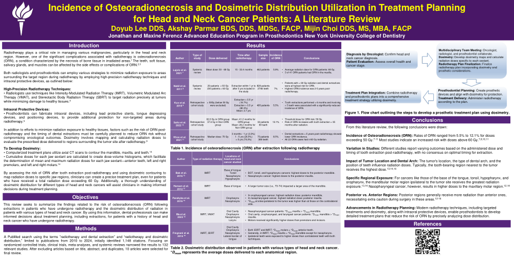
In addition to efforts to minimize radiation exposure to healthy tissues, factors such as the risk of osteoradionecrosis (ORN) post-radiotherapy and the timing of dental extractions must be carefully planned to reduce ORN risk without compromising oncological outcomes. Dosimetry involves mapping and calculating radiation doses to evaluate the prescribed dose delivered to regions surrounding the tumor site after radiotherapy.6
To Develop Dosimetry: