136 posters, 2 topics, 27 sessions, 339 authors, 88 institutions
ePostersLive by SciGen Technologies S.A. All rights reserved.
2024 ACP Annual Session
NOVEMBER 6-9, 2024 | MARCO ISLAND, FL JW MARRIOT

117
<em>Masticatory Function and Cognitive Health: Investigating a Molecular Connection Between Oral Tissues and the Brain</em>
Poster Presenter
Authors
Affiliations
Part of Topic
Original Research
Amongst the rapidly growing elderly population, there is increasing need for comprehensive prosthodontic treatment to address impaired masticatory function of many etiologies. Compromised oral health may contribute to a myriad of systemic pathologies, including cognitive decline. Several studies have observed an association between poor masticatory function in elderly patients and impaired cognitive function. Previously, we identified a novel retrograde transport mechanism of exosomal neprilysin, a neuroprotective enzyme, from the masseter muscle to the hippocampus through the trigeminal nerve.
Building on our previous studies suggesting that craniofacial tissue-derived exosomes may be delivered to the brain via retrograde axonal transport through cranial nerve junctions, we aim to further investigate this potential pathway. Specifically, this study will compare neurotrophic gene and protein expressions and their exosomal localizations between cell cultures of oral tissue and brain origins. Recent research has highlighted the importance of extracellular vesicles (EVs), particularly exosomes, in intercellular communication within the nervous system. Exosomes can carry a variety of cargo, including proteins, lipids, and nucleic acids, and have been shown to play roles in neuronal development, synaptic function, and neurological disorders.
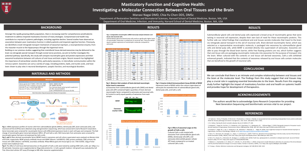
Fig 1. mRNA expression profiles of murine cells from submandibular-glands (WR21), dental-pulp (DP), brain-astrocytes (Ast), and neuronal cell line (CATHa) were obtained using next-generation-sequencing, which was outsourced to Zymo Research Corporation. Gene transcripts were screened for neurotrophic factors with differential expression levels in each cell types and compared. The fold change of neurotrophic gene transcripts between WR21, DP and Cath.a cells. NGS transcripts from astrocytes were screened for neurotrophic genes that were not expressed by Cath.a cells.
Fig 2. and 3. Brain-derived neurotrophic factor (BDNF) levels in exosomes and cell culture supernatant were analyzed via Western blot and ELISA, respectively. BDNF primary antibody mouse anti-BDNF (Abcam / ab108319), CD81 primary antibody, CD81 primary Ab: mouse anti-CD81 (Abcam / ab79559), secondary antibody: IRDye 800cw goat anti-mouse IgG (Li Cor / 926-32350). 10µg of total protein were loaded per lane.
Fig 4. The effect of oral tissue derived exosomes on the growth of Cath.a cells were tested by seeding 5000 Cath.acells per 100µL in a 96 well plate. Exosomes were supplemented at 20µg total protein/mL in Cath.a growth medium. Cell growth was evaluated by Cell Titer Glow total cellular ATP assay (Promega) at 48h after exosome supplementation
Fig 1. Comparative neurotrophic mRNA transcript analysis between oral tissue and neuronal cells
Submandibular gland and dental pulp cells produce significantly higher levels of neurotrophic molecules in comparison to neuronal cells. They also exhibit equal or greater expression of neurotrophic molecules compared to astrocytes.
Fig 2. Western blot analysis of brain-derived neurotropic factor level in exosomes
a) Exosomes from submandibular gland cells (SMG) and dental pulp cells (DP) contained higher quantities of brain-derived neurotrophic factor compared to astrocytes and neuronal cells. b) Control to verify equal quantity of exosomes from each sample
Fig 3. Enzyme-Linked Immunosorbent Assay (ELISA) of BDNF
Expression levels of BDNF within the culture supernatant of astrocytes far exceeded that of submandibular gland cells, dental pulp cells, and Cath.a cells
Fig 4. Effect of exosomal cargo on the growth of Cath.acells
Cultured Cath.a cells treated with exosomes from submandibular gland cells (SMG), dental pulp cells (DP), and astrocytes (Ast) exhibit greater cellular ATP levels compared to un-treated cultured Cath.a cells.
Submandibular gland cells and dental pulp cells expressed a broad array of neurotrophic genes that were lacking in neuronal cell expression, despite their own lack of need for these neurotrophic proteins. This further aids our initial findings that craniofacial and oral tissues secrete molecules that travel to the brain via retrograde axonal transport to the aid of neuronal cells. Brain-derived neurotrophic factor, which was selected as a representative neurotrophic molecule, is packaged into exosomes by submandibular gland cells and dental pulp cells, while BDNF is secreted directly into supernatant of astrocytes. Exosomes are able to pass through the blood brain barrier, while free floating proteins are not, which further suggests that oral tissue cells are packaging neurotrophic molecules into exosomes for the purpose of transport into the brain. When cultured with the contents or oral tissue cell derived exosomes, Cath.a cells exhibited enhanced growth, indicated that the contents of exosomes released by oral tissue cells contain molecules that are beneficial to the growth of neuronal cells.
We can conclude that there is an intimate and complex relationship between oral tissues and the brain at the molecular level. The findings from this study suggest that oral tissues may play a crucial role in supplying essential biomolecules to the brain. Results from this ongoing study further highlight the impact of masticatory function and oral health on systemic health and provides hope for development of therapeutics.