136 posters, 2 topics, 27 sessions, 339 authors, 88 institutions
ePostersLive by SciGen Technologies S.A. All rights reserved.
2024 ACP Annual Session
NOVEMBER 6-9, 2024 | MARCO ISLAND, FL JW MARRIOT

129
Impact of Palatal Height and Scanning Strategy on the Accuracy of Maxillary Denture IOS Digitization
Poster Presenter
Authors
Affiliations
Part of Topic
Original Research
Impact of Palatal Height and Scanning Strategy on the Accuracy of Maxillary Denture IOS Digitization
Sana Ghiba, Passent Ellakany, Kedith Sawangsri
Introduction: Different palatal anatomical configurations can often complicate the fabrication of complete denture prosthesis. Although intraoral scanning has been commonly used for complete denture duplication, some anatomical limitations, such as palatal configuration, may introduce errors. Previous studies have demonstrated that palatal configuration may have an effect on accuracy and speed of edentulous maxillary scans1,2.
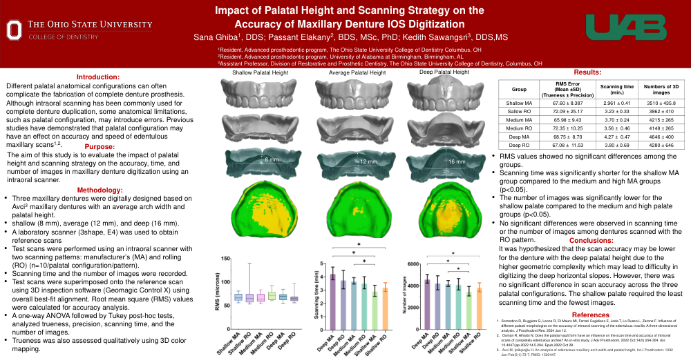
Purpose: To evaluate the impact of palatal height and scanning strategy on the accuracy, time, and number of images in maxillary denture digitization using an intraoral scanner.
Materials and Methods: Three maxillary dentures with varying palatal heights were digitally designed and printed: shallow (8 mm), average (12 mm), and high (16 mm). A laboratory scanner was used to obtain reference scans for each denture. Test scans were performed using an intraoral scanner with two scanning patterns: manufacturer’s (MA) and rolling (RO) (n=10). Scanning time and the number of images for each scan were recorded. Test scans were superimposed onto the reference scan using 3D inspection software for overall best-fit alignment. Root mean square (RMS) values were calculated for trueness analysis. A one-way Analysis of Variance (ANOVA), followed by Tukey post-hoc tests, analyzed trueness, scanning time, and the number of images. Trueness was also assessed qualitatively using 3D color mapping.
Results: The mean RMS of maxillary IOS digitization was 67.08 ±11.53 µm. RMS values showed no significant differences among the groups. Scanning time was significantly shorter for the shallow MA group compared to the medium and high MA groups (p<0.05). The number of images was significantly lower for the shallow palate compared to the medium and high palate groups (p<0.05). No significant differences were observed in scanning time or the number of images among dentures scanned with the RO pattern.
Conclusion: There was no significant difference in scan accuracy across the three palatal configurations. The shallow palate required the least scanning time and the fewest images.