127 posters, 2 topics, 24 sessions, 326 authors, 85 institutions
ePostersLive by SciGen Technologies S.A. All rights reserved.
2023 ACP ANNUAL SESSION
October 25-28 | San Diego, CA

73
Digitally Designed and Manufactured Sectional Custom Tray for a Patient with Microstomia
Poster Presenter
Authors
Marika Jagielska, Tanya Somohano-Marquez, Rafael Delgado-Ruiz
Affiliations
Part of Topic
Case Presentation
Digitally Designed and Manufactured Sectional Custom Tray for a Patient with Microstomia: A Case Report
Introduction
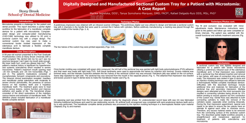
Microstomia presents a challenge for the patient and dental provider. This report describes a partial digital workflow for the fabrication of a mandibular complete denture for a patient with microstomia. Computer- aided design and computer-aided manufacturing (CAD-CAM) technology was utilized to 3D print a sectional custom tray with a unique design. The sectional custom tray was used to make a conventional border molded impression of the edentulous arch to fabricate a flexible complete mandibular denture.
Case Description
A 58-year-old woman presented to the Post Graduate Prosthodontics Clinic at Stony Brook University with a chief complaint “My dentist told me he can’t see me anymore because I can’t open my mouth wide enough. My lower teeth hurt, could you please help me?”. The patient’s medical history is significant for hypertension and systemic scleroderma which resulted in severe microsomia. Patient’s maximum mouth opening read ~20 mm vertically and ~35 mm horizontally (Figs. 1 and 2). The patient’s medications consisted of mycophenolate, lisinopril, omeprazole and oxycodone. The intraoral clinical examination revealed: maxillary and mandibular partial edentulism with moderate to severe chronic periodontitis, the presence of carious lesions on few maxillary teeth and 3 hopeless mandibular teeth. The treatment goals were to treat active carious lesions, restore function and improve esthetics and quality of life. The transitional phase consisted of the direct restorations of the maxillary carious lesions and extraction of mandibular teeth. The definitive phase included fabrication of a mandibular flexible complete denture.
Technique/Photos
A preliminary impression was obtained with an intraoral scanner (3Shape). The preliminary digital cast was utilized to design and fabricate a sectional custom tray with 2 channels in the handle that were used to connect the 2 halves with cylinders. Before additively manufacturing, a marking was placed through the sagittal middle of the handle (Figs. 3, 4).
The two halves of the custom tray were printed separately (Figs. 5,6).
Once border molding was completed with green stick compound, the left half of the sectional tray was painted with light body polyvinylsiloxane (PVS) adhesive and final wash was made with light body PVS. Soft tissue landmarks were used to approximate the halves for insertion and removal. Excess material was trimmed away, and the intimate connection between the two halves of the sectional custom tray was ensured. Petroleum jelly was added on the cut surface. Same step repeated for right side. The sectional tray was removed from the mouth in two separate pieces (Fig. 7). The obtained final impression was beaded and boxed, poured in type III dental stone to make the mandibular master cast (Fig. 8).
An opposing cast was printed from the intraoral scan obtained during the diagnostic phase. An underextended record base with wax rim was fabricated following traditional techniques and used for jaw relationship records. An artificial tooth arrangement was completed with semi-anatomical denture teeth and a try in was performed. The mandibular complete dental prosthesis was processed by the injection molding technique in a thermoplastic flexible nylon material (Valplast) (Fig. 9) and inserted.
The fit and occlusion was evaluated with minor adjustments made. Maintenance instructions were given to the patient and follow ups were completed at timely intervals. The patient was satisfied with the overall functional and esthetic treatment outcome (Fig. 10).
Conclusions
A sectional custom tray was digitally designed and fabricated for a patient with severe microstomia utilizing a preliminary intraoral scan of the edentulous mandibular arch. A final impression was completed with a sectional tray that allowed insertion and removal in two halves, with ease of connection intra and extra orally. The impression was casted in type III stone and an injection molding technique was utilized to fabricate a thermoplastic flexible complete mandibular denture. Difficulties with the described technique include additional time and materials for fabrication of the sectional tray and secondary impression. Multiple designs were attempted and the most successful one resulted in connecting cylinders being shrunk by 10% before printing to ensure passive but firm connection of the custom tray. Dental floss was attached to both connecting cylinders to make the removal of the cylinders easier, especially when working intraorally. During the final impression appointment, special care needs to be taken to ensure the two pieces have an intimate fit against each other to minimize errors. Therefore, any excess material needs to be removed from the connecting surface of the sectional custom tray. The described partial digital workflow presents a straightforward and inexpensive approach for treatment of patients with scleroderma and microstomia.