127 posters, 2 topics, 24 sessions, 326 authors, 85 institutions
ePostersLive by SciGen Technologies S.A. All rights reserved.
2023 ACP ANNUAL SESSION
October 25-28 | San Diego, CA

179
Treatment Planning for Class III Malocclusion with Anterior/Posterior Open Bite: A Case Report
Poster Presenter
Authors
Affiliations
Part of Topic
Case Presentation
Introduction:
Class III malocclusion, characterized by maxillary retrognathism or mandibular prognathism, often results in anterior teeth adopting an end-to-end relationship¹, causing functional and aesthetic challenges. Traditional treatment involves orthodontic intervention in addition to orthognathic surgery to establish a Class I occlusal relationship.² This case report presents a 42-year-old male with Class III occlusion and anterior/posterior open bite. The patient's main concerns were impaired biting, speech difficulties, and aesthetic concerns. In response, an innovative and less invasive approach was proposed, bypassing surgical and orthodontic interventions. This approach utilizes single-unit fixed prostheses to restore occlusion while maintaining the visual appearance of an end-to-end anterior dentition. While offering a less invasive alternative, this approach necessitates a comprehensive evaluation, particularly regarding its impact on speech, including closest speaking space and articulation of sounds.
A review of the patient’s medical history revealed hypertension controlled by Hydrochlorothiazide and a previous gastric bypass surgery resulting in a chronic pain condition managed by Trazodone, Topiramate, Pregabalin, Zofran, and Ambien. Also included in the medical history was a severe unknown illness around the age of 18 months that was treated using various undefined antibiotics in Nigeria. The patient denied any history of smoking or alcohol consumption. The patient also denied previous orthodontic treatment.
Clinical Assessment:
An initial appointment consisting of a full mouth series of radiographs, a comprehensive exam, diagnostic impression making, and photographs was performed. The radiographic examination revealed fully-erupted third molars in the upper right, lower left, and lower right quadrants, along with bilateral missing mandibular first molars supported by previous radiographs indicating caries-related tooth loss. Interproximal composite restorations and fixed-single-unit ceramic prostheses on maxillary anterior teeth, done for esthetic purposes a decade ago, were noted. It was also noted that the restoration of #6 is missing. Generalized bone loss ranging from 0% to 15% was observed. The comprehensive examination confirmed these findings.
Additionally, an occlusal analysis revealed the presence of both anterior and posterior open bite, with occlusal contacts restricted solely to the second and third molars. Furthermore, minimal occlusal wear was observed during the examination. The periodontal examination showed probing depths ranging from 1-6 and CAL ranging from 0-5. The gingival tissues were noted to be cyanotic, edematous, and bulbous. The intraoral and extraoral cancer screening were within normal limits and there were no significant findings upon examining the temporomandibular joint.
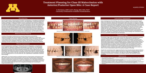
Upon reviewing the clinical findings, the patient was diagnosed with Generalized Stage II Grade A Periodontitis³, anterior open bite, posterior open bite, Class III occlusion, a missing restoration, and partial edentulism. Following the initial examination, the diagnostic models were mounted on an articulator and analyzed. The casts were duplicated and cross-mounted, followed by a simulated occlusal equilibration which revealed an exacerbation of the Class III malocclusion and resulted in an underbite. Subsequently, the casts were returned to their original positions, and a diagnostic wax-up was completed. A transfer putty was created to transfer the wax-up into the patient's oral cavity, utilizing Protemp provisional crown material. A comprehensive series of tests ensued to evaluate both speech functionality and esthetic outcomes. The patient reported enhanced enunciation and an overall improved appearance. Taking into consideration the previous clinical findings and confirmation of the wax-up, a prosthetic treatment plan was developed.
The primary stage of the treatment plan involved the disease phase, developed in response to the periodontal diagnosis. This phase consists of scaling and root planing all four quadrants, followed by a re-evaluation. The second phase of treatment is focused on the definitive prostheses. The loss of mandibular first molars due to caries and the presence of demarcated enamel chroma on the mandibular incisors, coupled with the patient's history of severe illness during infancy, indicates a developmental condition known as molar-incisor hypomineralization.⁴ To restore function and aesthetics, the two missing molars will be replaced with endosteal implants, and full-coverage crowns will be placed on teeth #3-14 and #20-29 to create occlusal contact. Considering the hypomineralized enamel on the mandibular incisors, a decision was made to opt for full-coverage crowns over veneers as there is a lack of mineralized enamel of the planned margin for bonding.
Despite the conventional ortho-surgical method for Class III malocclusion, fixed prosthetics offer a non-invasive solution to address the patient’s complaints of impaired biting, speech issues, and aesthetic concerns related to the open bite. This approach highlights the versatility of prosthodontic techniques, providing an efficient and minimally invasive resolution.
-
Zere E, Chaudhari PK, Sharan J, Dhingra K, Tiwari N. Developing Class III malocclusions: challenges and solutions. Clin Cosmet Investig Dent. 2018 Jun 22;10:99-116.
-
Behnia H, Mohammad-Rahimi H, Behnaz M. Treatment of an Adult Skeletal Class III Patient with Surgically Assisted Rapid Palatal Expansion and Facemask. Case Rep Dent. 2019 Dec 30;2019:8251903.
-
Sanz M, Herrera D, Kebschull M, Chapple I, Jepsen S, Beglundh T, Sculean A, Tonetti MS; EFP Workshop Participants and Methodological Consultants. Treatment of stage I-III periodontitis-The EFP S3 level clinical practice guideline. J Clin Periodontol. 2020 Jul;47 Suppl 22(Suppl 22):4-60. doi: 10.1111/jcpe.13290. Erratum in: J Clin Periodontol. 2021 Jan;48(1):163.
-
Krishnan R, Ramesh M. Molar incisor hypomineralisation: A review of its current concepts and management. SRM J Res Dent Sci. 2014;5:248–52.