127 posters, 2 topics, 24 sessions, 326 authors, 85 institutions
ePostersLive by SciGen Technologies S.A. All rights reserved.
2023 ACP ANNUAL SESSION
October 25-28 | San Diego, CA

161
3D-Facial Scanning for Diagnosis of Ideal Smile Designs and Occlusion
Poster Presenter
Authors
Affiliations
DUMMY, DUMMY2, Nova Southeastern University Postgraduate Prosthodontics
Part of Topic
Case Presentation
Introduction
Three-dimensional facial scanning has given way to facially-driven prosthodontic treatment. Historically, the origins of facial scanning developed from the government and civil authority sector. Today, it has become a valuable diagnostic tool in dentistry which allows for evaluation of all possible esthetic and occlusion parameters simultaneously, while creating a customized smile design. Thus, by directly incorporating the patient’s face, the customized smile design is not simply “prosthetically-driven.” Rather, the entire course of treatment can now be termed as: “facially-driven.” At Nova Southeastern University, a 3-D facial scanner (RAYFace, Ray America) is currently used to enhance prosthodontic treatment planning in complex and esthetically demanding oral rehabilitation cases. A case presentation which utilized a facial scan for diagnosis and treatment planning will be featured.
Methods and Techniques
A seventy-nine-year-old male presented to the NSU Postgraduate Prosthodontics Clinic for a full mouth rehabilitation. The patient had a worn dentition which indicated the need to restore the vertical dimension of occlusion (OVD). Composite was placed on teeth #22 and #27 to arbitrarily restore the OVD for diagnostic purposes. The following records were collected at the patient’s initial visit: (1) maxillary, mandibular, and bite intraoral scans (with composite used to hold the new OVD position), (2) 3-D facial scan, (3) full CBCT (including cranium).
The 3-D facial scan was recorded using a facial scanner which utilized six cameras to capture the patient’s face. Additional cameras built into the scanner focused solely on capturing the anterior dentition region. The patient was positioned to ensure his head was not tilted, his gaze was straight on, and his lips were aligned to the dentition region outlined in the camera guidelines. The patient was then instructed to smile as the facial images were swiftly acquired within 2-3 seconds and automatically gathered by the scanner software to produce a 3-D facial reconstruction.
The intraoral scans and CBCT records were imported into the proprietary facial scanner software where all data and records were seamlessly merged (Figure 1). The initial alignment was then directly integrated into a third-party smile design software to complete the diagnostic wax-up.
FIGURES
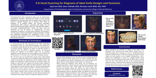
Figure 1: Ability for Multiple Data Set Integration The software began by aligning the maxillary, mandibular, and bite intraoral scans. The CBCT was then aligned to maxillary intraoral scan. The third and final layer, the facial scan, was merged.
Figures 2a &2b: Frontal and Profile Views of Integration This alignment was confirmed by using the proper location of the condyles and infra-orbital lines as they align the CBCT scan directly to the facial scan.
Figure 3: Facial Guide Planes Next, the following facial lines and angles were prompted and initially set by the software: midline, interpupillary, incisal, ala-tragus, horizontal Frankfurt plane, SNA/FMA/FMR angles. These facial lines and angles were reviewed in correlation with the facial scan, CBCT, and intraoral scans, and deemed to be in the correct position.
Figure 4a & 4b: Virtual Articulation Integration in Smile Design Software Finally, the projected file directly transferred from the proprietary software into a third-party smile design software. This direct integration allowed for all the patient records to remain accurately stitched together as well as an accurate digital facebow articulation in the smile design software.
Figures 5a & 5b: Facially-Driven Digital Smile Design A diagnostic smile design was created by a lab technician and reviewed, modified, and approved by the clinician.
Discussion
From the initial alignment, it was clear that the facial midline and dental midline would not coincide. However, this was within acceptable limits and can be adjusted by the clinician. Furthermore, the excessive flaring of the anterior teeth was easily diagnosed in the profile view of Figure 2b.
While creating the diagnostic smile design, the lab technician was simultaneously able to evaluate facial lines and lip position against the teeth position, incisal edge, shape, width, length, and occlusal plane. After receiving the diagnostic design, the clinician was able to confirm the esthetics and occlusion of the smile design and determine the necessary teeth preparations. Elective endodontic therapy was applied on several teeth which required additional preparation. After confirmation of the diagnostic smile design, a diagnostic wax-up model was printed and full arch maxillary and mandibular provisional shells were milled in PMMA.
Conclusion
Dental restorations are constantly on display in the center of an individual’s face. Therefore, the dental industry should focus on achieving facially-driven treatment as well as prosthetically-driven treatment. With the advent of 3-D facial scanning, all aspects of smile design and tooth position can be evaluated simultaneously with a patient’s face. Furthermore, it allows for a highly accurate virtual articulation with the TMJ joint position from the CBCT. This is of great value not only to the clinician, but also to the communicating lab technician. Using a 3-D facial scan for diagnosis and treatment planning will save time and try-in visits, as well as positional and tooth selection errors that may occur without this technology.
REFERENCES
1. Three-Dimensional Facial Scanning: A New Paradigm. Vafiadis D. Dentistry Today. 2023, 69-73.
2. Facial Scanners in Dentistry: An Overview. Lee JD, Nguyen O, Lin Y-C, et al. Prosthesis. 2022; 4(4):664-78.
3. 3D Facial Scanning: Dual-Arch Fixed Prosthesis From Diagnosis to Delivery. Bynum JH, Mirelez JA. Compend Contin Educ Dent. 2021;42(5):230-5.
4. Comparison of 3D Scanning Versus 2D Photography for the Identification of Facial Soft-Tissue Landmarks. Zogheib T, Jacobs R, Bornstein MM, et al. Open Dent J. 2018;31(12):61-71.