127 posters, 2 topics, 24 sessions, 326 authors, 85 institutions
ePostersLive by SciGen Technologies S.A. All rights reserved.
2023 ACP ANNUAL SESSION
October 25-28 | San Diego, CA

138
Pickup Impression Technique to Facilitate Digital Merge of Patient Provisional Restorations with Cone Beam Computed Tomography Scan
Poster Presenter
Authors
Affiliations
DUMMY, Boston University Henry M. Goldman School of Dental Medicine
Part of Topic
Case Presentation
Pickup Impression Technique to Facilitate Digital Merge of Patient Provisional Restorations with Cone Beam Computed Tomography Scan
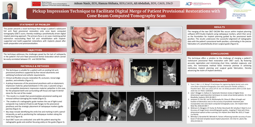
This poster presents a novel technique that merges a patient's radiolucent full arch fixed provisional restoration onto cone beam computed tomography (CBCT) scans, thereby enabling a prosthetically driven digital implant planning approach. The primary focus is on cases involving partial edentulism necessitating fixed full arch rehabilitation with implant-supported and tooth-supported restorations with implants placed after tooth preparation and provisionalization.
The technique addresses the challenges posed by the lack of radiopacity in the patient's full arch fixed provisional dental restoration which cannot be easily correlated between STL and DICOM files.
The merging of the two CBCT DICOM files occurs within implant planning software (DTX Studio Implant) using radiopaque markers, which then serve as the foundation for implant planning guided by the provisional teeth position. The results underscore the successful alignment of radiographic data with the patient's provisional teeth position, culminating in the precise fabrication of a prosthetically driven surgical guide (Figure 6).
This technique offers a solution to the challenge of merging a patient's radiolucent provisional fixed restoration with CBCT scans. By fostering accurate registration and minimizing chair time, radiation exposure, and costs, this noninvasive method holds immense promise in achieving meticulous implant planning and surgical guide fabrication, thereby advancing the realm of implant dentistry.