127 posters, 2 topics, 24 sessions, 326 authors, 85 institutions
ePostersLive by SciGen Technologies S.A. All rights reserved.
2023 ACP ANNUAL SESSION
October 25-28 | San Diego, CA

121
Fabrication of a zygomatic implant-retained obturator utilizing digital workflow: A Case Report
Poster Presenter
Authors
Evangelia Gkoulitou, Noora Almasoodi, Ahmad Reza Edalati Sharbaf, Jorge Palavicini
Affiliations
Part of Topic
Case Presentation
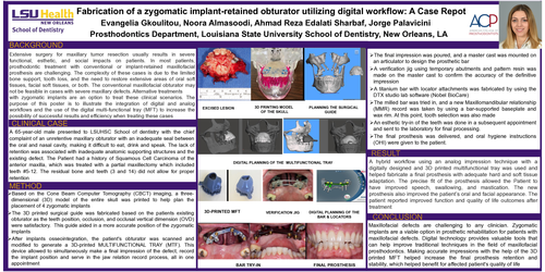
CLINICAL CASE: A 65-year-old male presented to LSUHSC School of dentistry with the chief complaint of an unretentive maxillary obturator with an inadequate seal between the oral and nasal cavity, making it difficult to eat, drink and speak. The lack of retention was associated with inadequate anatomic supporting structures and the existing defect. The Patient had a history of Squamous Cell Carcinoma of the anterior maxilla, which was treated with a partial maxillectomy which included teeth #5-12. The residual bone and teeth (3 and 14) did not allow for proper retention
METHOD: 1. Based on the Cone Beam Computer Tomography (CBCT) imaging, a three- dimensional (3D) model of the entire skull was printed to help plan the placement of 4 zygomatic implants. 2. The 3D printed surgical guide was fabricated based on the patients existing obturator as the teeth position, occlusion, and occlusal vertical dimension (OVD) were satisfactory. This guide aided in a more accurate position of the zygomatic implants. 3. After implants osseointegration, the patient's obturator was scanned and modified to generate a 3D-printed MULTIFUNCTIONAL TRAY (MTF). This device allowed to simultaneously make a final impression of the defect, record the implant position and serve in the jaw relation record process, all in one appointment. 4. The final impression was poured, and a master cast was mounted on an articulator to design the prosthetic bar. 5. A verification jig using temporary abutments and pattern resin was made on the master cast to confirm the accuracy of the definitive impression. 6. A titanium bar with locator attachments was fabricated by using the DTX studio lab software (Nobel BioCare). 7. The milled bar was tried in, and a new Maxillomandibular relationship (MMR) record was taken by using a bar-supported baseplate and wax rim. At this point, tooth selection was also made. 8. An esthetic try-in of the teeth was done in a subsequent appointment and sent to the laboratory for final processing. 9. The final prosthesis was delivered, and oral hygiene instructions (OHI) were given to the patient.
RESULT: A hybrid workflow using an analog impression technique with a digitally designed and 3D printed multifunctional tray was used and helped fabricate a final prosthesis with adequate hard and soft tissue adaptation. The precise fit of the prosthesis allowed the Patient to have improved speech, swallowing, and mastication. The new prosthesis also improved the patient’s oral and facial appearance. The patient reported improved function and quality of life outcomes after treatment
CONCLUSION: Maxillofacial defects are challenging to any clinician. Zygomatic implants are a viable option in prosthetic rehabilitation for patients with maxillofacial defects. Digital technology provides valuable tools that can help improve traditional techniques in the field of maxillofacial prosthodontics. Making accurate impressions with the help of the 3D printed MFT helped increase the final prosthesis retention and stability, which helped benefit for affected patient's quality of life