105 posters, 2 topics, 24 sessions, 264 authors, 71 institutions
ePostersLive by SciGen Technologies S.A. All rights reserved.
2022 ACP Annual Session
2 - 5 November | Austin, TX

77
Fully digital workflow for fabrication of a 3D-printed ear stent for Auricular Keloid:
Poster Presenter
Authors
Ahmad Reza Edalati Sharbaf, Amir Nejat, Soleiman Hamdan, Isaias Abrego, John Lindsey, Roger Vitter
Affiliations
Part of Topic
Case Presentation
Fully Digital Workflow for Fabrication of A 3D Printed Ear Stent for Auricular Keloids
Ahmad Reza Edalati, Amir H Nejat, Suleiman Hamdan, Isaias Abrego, John T Lindsey, Roger Vitter
Prosthodontics Department, LSUHSC School of Dentistry, New Orleans, LA
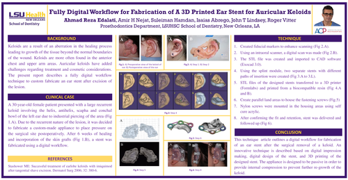
BACKGROUND:Keloids are a result of an aberration in the healing process leading to growth of the tissue beyond the normal boundaries of the wound. Keloids are more often found in the anterior chest and upper arm areas. Auricular keloids have added challenges regarding treatment and cosmetic considerations. The present report describes a fully digital workflow technique to custom fabricate an ear stent after excision of the lesion.
CLINICAL CASE
A 30-year-old female patient presented with a large recurrent keloid involving the helix, antihelix, scapha and conchal bowl of the left ear due to industrial piercing of the area (Fig 1.A). Due to the recurrent nature of the lesion, it was decided to fabricate a custom-made appliance to place pressure on the surgical site postoperatively. After 6 weeks of healing and incorporation of the skin grafts fig (1.B), a stent was fabricated using a digital workflow.
TECHNIQUE
CONCLUSION
This technique article outlines a digital workflow for fabrication of an ear stent after the surgical removal of a keloid. An innovative technique is described based on digital impression making, digital design of the stent, and 3D printing of the designed stent. The appliance is designed to be passive in order to provide internal compression to prevent further re-growth of the keloid.