105 posters, 2 topics, 24 sessions, 264 authors, 71 institutions
ePostersLive by SciGen Technologies S.A. All rights reserved.
2022 ACP Annual Session
2 - 5 November | Austin, TX

100
Rebirth of the Mucostatic Impression in the Digital Era
Poster Presenter
Authors
Affiliations
Part of Topic
Case Presentation
Introduction:
When fabricating a complete removable prosthesis it is important to make sure that it is not only esthetically acceptable, but also functionally acceptable. The bone underlying a removable prosthesis can affect the fit and overall function. In cases where there has been resorption of the bone the denture can have less stability and it can compromise the effectiveness of the denture. The purpose of this poster is to present a patient case where digital technology was used as part of a mucostatic impression and to fabricate a maxillary complete denture with a metal base.
Clinical Report:
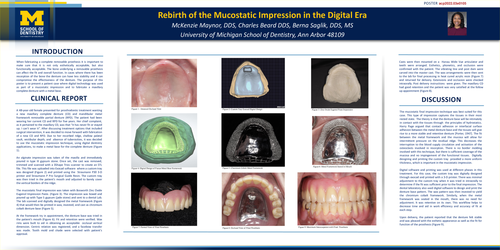
A 48-year-old female presented for prosthodontic treatment wanting a new maxillary complete denture (CD) and mandibular metal framework removable partial denture (RPD). The patient had been wearing her current CD and RPD for five years. Her chief complaint, as it pertained to the maxillary CD, was that “it has never fit or stayed up. I can’t wear it”. After discussing treatment options that included surgical intervention, it was decided to move forward with fabrication of a new CD and RPD. Due to her resorbed ridge, shallow palatal vault, vestibular depth, and absence of tuberosities, it was decided to use the mucostatic impression technique, using digital dentistry applications, to make a metal base for the complete denture (Figure 1).
An alginate impression was taken of the maxilla and immediately poured in type III gypsum stone. Once set, the cast was removed, trimmed and scanned with a 3Shape Triosscanner to create an STL file. This file was uploaded into Exocad software where a custom tray was designed (Figure 2) and printed using the Straumann P30 3-D printer and Straumann P Pro Surgical Guide Resin. The custom tray was then tried in the patient's mouth and adjusted to barely cover the vertical borders of the ridge.
The mucostatic final impression was taken with Bosworth Zinc Oxide Eugenol Impression Paste. (Figure 3). The impression was boxed and poured up with Type 5 gypsum (jade stone) and sent to a dental Lab. The lab scanned and digitally designed the metal framework (Figure 4) that would then be printed in wax, invested, and cast as chromium cobalt denture base (Figure 5).
At the framework try in appointment, the denture base was tried in the patient's mouth (Figure 6). Fit and retention were verified. Wax rims were built to aid in obtaining an acceptable occlusal vertical dimension. Centric relation was registered, and a facebow transfer was made. Tooth mold and shade were selected with patient’s approval.
Casts were then mounted on a Hanau Wide Vue articulator and teeth were arranged. Esthetics, phonetics, and occlusion were confirmed with the patient. The vibrating line and post dam were carved into the master cast. The wax arrangements were then sent to the lab for final processing in heat cured acrylic resin (Figure 7) and returned for delivery. Extensions and occlusion were checked intraorally. Post delivery instructions were given. The maxillary CD had good retention and the patient was very satisfied at the follow up appointment (Figure 8).
Discussion:
The mucostatic final impression technique was best suited for this case. This type of impression captures the tissues in their most rested state. The theory is that the denture base will be intimately in contact with the tissues through the principles of hydrostatics. Harry Page argued that contact adhesion or interfacial surface adhesion between the metal denture base and the tissues will give rise to a more stable and retentive denture [Porter, 1947]. The fit between the metal framework and the mucosa will allow for intermittent pressure on the residual ridge. This decreases the interruption to the blood supply circulation and activation of the osteoclasts involved in resorption. There is no border molding involved with this technique, but there is sufficient coverage of the mucosa and no impingement of the functional tissues. Digitally designing and printing the custom tray provided a more uniform thickness, which is important in the mucostatic impression.
Digital software and printing was used at different phases in this treatment. For this case, the custom tray was digitally designed through exocad and printed with a 3-D printer. There was minimal adjustment to the custom tray when it was tried in intraorally to determine if the fit was sufficient prior to the final impression. The dental laboratory also used digital software to design and print the denture base pattern. The wax pattern was then invested to yield the chromium cobalt framework. Similarly, when the metal framework was seated in the mouth, there was no need for adjustment. It was retentive on its own. This workflow helps to decrease time and aid in work efficiency and accuracy of fit at each step.
Upon delivery, the patient reported that the denture felt stable and was pleased with the esthetic appearance as well as the fit for function of the prosthesis (Figure 9).