105 posters, 2 topics, 24 sessions, 264 authors, 71 institutions
ePostersLive by SciGen Technologies S.A. All rights reserved.
2022 ACP Annual Session
2 - 5 November | Austin, TX

60
In Vitro 3-Dimensional Comparison of Two Digital Workflows For CAD/CAM Immediate Dentures: A Pilot Study
Poster Presenter
Part of Topic
Case Presentation
In Vitro 3-Dimensional Comparison of Two Digital Workflows For CAD/CAM Immediate Dentures: A Pilot Study
Nahyun Lee DDS MS, Thomas Giugliano DDS, Mijin Choi DDS MS MBA FACP
Jonathan and Maxine Ferencz Advanced Education Program in Prosthodontics, New York University College of Dentistry
INTRODUCTION
The utilization of computer-aided design and computer-aided manufacturing (CAD-CAM) technology in dentistry is expanding as the technologies advance. Digital dentistry can improve treatment efficiency by reducing fabrication time in the dental laboratory. CAD/CAM technology was introduced to fabricate complete dentures in 1994.1 Digital denture technique allows for shortened chair time, improved denture retention, less polymerization shrinkage of the denture base, and easy duplication of dentures.2 However, intraoral scanners are ineffective in capturing functional border extensions for removable prostheses.
OBJECTIVE
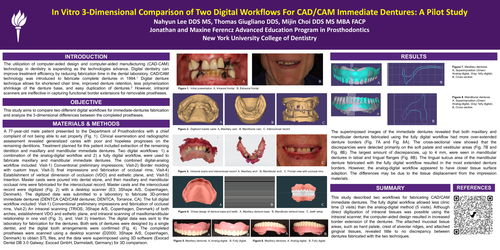
This study aims to compare two different digital workflows for immediate-dentures fabrication and analyze the 3-dimensional differences between the completed prostheses.
MATERIALS & METHODS
A 77-year-old male patient presented to the Department of Prosthodontics with a chief complaint of not being able to eat properly (Fig. 1). Clinical examination and radiographic assessment revealed generalized caries with poor and hopeless prognoses on the remaining dentitions. Treatment planned for this patient included extraction of the remaining dentition and maxillary and mandibular immediate dentures. Two digital workflows: 1) a combination of the analog-digital workflow and 2) a fully digital workflow, were used to fabricate maxillary and mandibular immediate dentures. The combined digital-analog workflow included: Visit-1) Conventional preliminary impressions; Visit-2) Border molding with custom trays; Visit-3) final impressions and fabrication of occlusal rims; Visit-4) Establishment of vertical dimension of occlusion (VDO) and esthetic plane, and; Visit-5) Insertion. Master casts were poured into dental stone, and then maxillary and mandibular occlusal rims were fabricated for the interocclusal record. Master casts and the interocclusal record were digitized (Fig. 2) with a desktop scanner (E3; 3Shape A/S, Copenhagen, Denmark). The digitized data was submitted to a laboratory to fabricate 3D-printed immediate dentures (DENTCA CAD/CAM dentures; DENTCA, Torrance, CA). The full digital workflow included: Visit-1) Conventional preliminary impressions and fabrication of occlusal rims, Visit-2) An intraoral scanning (TRIOS; 3Shape A/S, Copenhagen, Denmark) of both arches, establishment VDO and esthetic plane, and intraoral scanning of maxillomandibular relationship in one visit (Fig. 3), and; Visit 3) Insertion. The digital data was sent to the laboratory for fabrication for the dentures. Both sets of dentures were designed by a single dentist, and the digital tooth arrangements were confirmed (Fig. 4). The completed prostheses were scanned using a desktop scanner (D2000; 3Shape A/S, Copenhagen, Denmark) to obtain STL files, and the data were superimposed using 3D software (Exocad Dental DB 3.0 Galway; Exocad GmbH, Darmstadt, Germany) for 3D comparison.
RESULTS
The superimposed images of the immediate dentures revealed that both maxillary and mandibular dentures fabricated using the fully digital workflow had more over-extended denture borders (Fig. 7A and Fig. 8A). The cross-sectional view showed that the discrepancies were detected primarily on the soft palate and vestibular areas (Fig. 7B and Fig. 8B). The largest amount of discrepancies, up to 4 mm, were seen in mandibular dentures in labial and lingual flanges (Fig. 8B). The lingual sulcus area of the mandibular denture fabricated with the fully digital workflow resulted in the most extended denture borders. However, the analog-digital workflow appeared to have closer tissue surface adaption. The differences may be due to the tissue displacement from the impression materials.
SUMMARY
This study described two workflows for fabricating CAD/CAM immediate dentures. The fully digital workflow allowed less clinic time (3 visits) than the analog-digital method (5 visits). Although direct digitization of intraoral tissues was possible using the intraoral scanner, the computer-aided design resulted in increased border extension of the dentures. The attached mucosal tissue areas, such as hard palate, crest of alveolar ridges, and attached gingival tissues, revealed little to no discrepancy between dentures fabricated with the two techniques.