105 posters, 2 topics, 24 sessions, 264 authors, 71 institutions
ePostersLive by SciGen Technologies S.A. All rights reserved.
2022 ACP Annual Session
2 - 5 November | Austin, TX

134
A Novel approach to fabricating Surgical, Interim and Definitive obturator fabrication
Poster Presenter
Authors
Affiliations
Part of Topic
Case Presentation
A novel approach to CAD/CAM Surgical, Interim, and Definitive Obturator fabrication
Introduction
The execution of prosthetic rehabilitation for patients with oral cancer post-surgical intervention has been revolutionized with the use of digital technology in scanning, designing, and printing of final prostheses. This has allowed prosthodontists and surgeons to work more coherently in planning, prior to surgical intervention and executing of reconstructive cases to achieve a more ideal result. The purpose of this poster presentation is to illustrate a digital workflow in reconstruction of a patient following partial maxillectomy.
Case Report
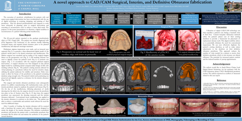
The 82-year-old patient reported to the graduate prosthodontic clinic at UNC Chapel Hill. The patient was recently diagnosed with pT4a Squamous Cell Carcinoma of the anterior maxilla following excisional biopsy and required dental clearance prior to his planned maxillectomy and adjuvant oncologic treatment.
Preliminary alginate impressions were made, and an intraoral scan captured (Fig. 5), to generate a three-dimensional digital model of the patient’s mouth prior to his dental extractions and maxillectomy. The ablative surgeon was consulted, and an outline of the surgical margins drawn on the preoperative cast (Fig. 2). A 3D meshing software was used to digitally extract the patient’s teeth (Fig. 6) to perform cast surgery (Fig. 7) in accordance with the surgeon’s planned surgical intervention. A surgical obturator was then designed on this altered digital model by combining dental computer-aided design (CAD) with meshing software (Fig. 8). The patient’s existing anatomy allowed for virtual planning of a prosthesis without the need for retentive clasps, utilizing the palatal undercuts for stability and retention. Holes were made to allow for fixture of the prosthesis to the patient’s remaining dentition using wrought wire. The patient’s initial tooth positioning was preserved and digitally copied as a reference for the interim obturator (Figs. 9 & 10).
The surgical and interim obturator prostheses were subsequently printed via additive manufacturing with methylacrylate resin, and the interim prosthesis stained to replicate the patient’s soft tissues (Fig 12). On the day of the maxillectomy, following placement of sterile dressing into the defect site, the surgeon elected to screw the prosthesis into place in lieu of fixation wires (Fig. 4). 21 days later, the surgical prosthesis was unscrewed, the dressing removed, and the interim obturator delivered. Conventional tissue conditioning was applied to the interim obturator to conform to the defect site. The patient was able to achieve a comfortable and aesthetic result without the need the need for additional clasps.
After 3 months of healing, the interim obturator will be scanned with a benchtop lab scanner and a definitive obturator designed to mimic the design of the interim obturator. The definitive monoblock prosthesis will provide the patient with a comfortable prosthesis that accurately fills the defect site and mimics the aesthetics of the patient’s natural dentition.
Discussion
Utilization of digital CAD/CAM technology can help expediate a patient’s care during a traumatic time in their lives. Current baseplate fabrication methods utilize the patient’s plaster cast of their existing dentition to provide a framework for placement of replacement denture teeth. This digital workflow allows us to potentially eliminate the need for conventional impressions. We can now effectively duplicate the patient’s existing dentition, as well as make minor adjustments to the dentition on their definitive obturator prostheses. This approach reduces the number of visits needed for a patient to receive their definitive prosthesis, which in turn increase our clinical efficiency. The potential for increased patient comfort and the reduced number of postop appointments.
Acknowledgement
The authors would like to thank Dewey Chapa, Lead Digital Dental Technician, for his assistance with the CAD Laboratory phase; UNC’s Maxillofacial prosthetic section. The authors reported no conflicts of interested related to this case report.
Reference
1. Soltanzadeh, Pooya, et al. “Obturator fabrication incorporating computer-aided design and 3-dimenstional printing technology: a clinical report.” The Journal of Prosthetic Dentistry 121.4 (2019): 694-697
2. Londono, Jimmy, et al. "Fabrication of a definitive obturator from a 3D cast with a chairside digital scanner for a patient with severe gag reflex: a clinical report." The Journal of Prosthetic Dentistry 114.5 (2015): 735-738.
3. Park, Jin-Hong, et al. "Fabricating a Maxillary Obturator Using an Intraoral Digital Impression: A Case History Report." International Journal of Prosthodontics 30.3 (2017).