66 posters, 2 videos, 1 audios, 2 topics, 162 authors, 49 institutions
ePostersLive by SciGen Technologies S.A. All rights reserved.
American College of Prosthodontists - 2020 ANNUAL SESSION
November 4-7 | Virtual Meeting

80
Fabrication of a floating implant and bone surgical guide using digital technology: A technique article
Poster Presenter
Authors
Affiliations
Part of Topic
Case Presentation
Fabrication of an Innovative Surgical Guide Using Digital Technology: A Technique Report
Computer-aided design and computer-aided manufacturing (CAD-CAM) technologies have contributed to guided implant surgery.1CAD-CAM surgical guides can be tooth, bone, or mucosa supported. According to some studies, bone-supported guides can be very accurate. Tissue supported guides are also accurate but if errors occur during surgery it is attributable to mucosal compressibility.2,3However, the inaccuracies encountered with bone supported guides are due to inherent errors with the cone beam computed tomography (CBCT) unit and the implant planning software.4,5,6,7Bone Supported guides can be used for either bone reduction or implant placement; Bone supported implant guides following bone reduction can be designed in various ways. One way is to design the guide to fit directly over the reduced bone surface (like the guide presented in this report). Another way is to design the guide to be stacked over the bone reduction guide. To minimize inaccuracies, a technique is presented in this report to create a bone supported guide that engages only the buccal bone and is independent of any precedented bone reduction as compared to the stacked-type guide.
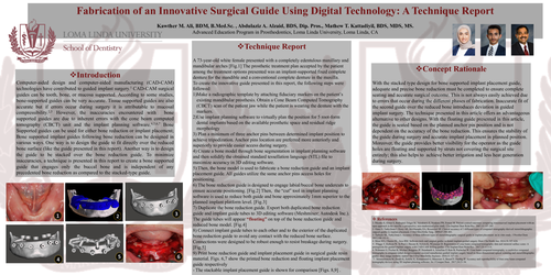
A 73-year-old white female presented with a completely edentulous maxillary and mandibular arches [Fig.1] The prosthetic treatment plan accepted by the patient among the treatment options presented was an implant-supported fixed complete denture for the mandible and a conventional complete denture in the maxilla.
To create the innovative guide presented in this report, the following steps were followed:
3) Plan a minimum of three anchor pins between determined implant position to achieve tripodization. Anchor pins location are preferred more anteriorly and superiorly to provide easier access during surgery.
4) Create a bone model through bone segmentation in implant planning software and then solidify the obtained standard tessellation language (STL) file to maximize accuracy in 3D editing software.
5) Then, the bone model is used to fabricate a bone reduction guide and an implant placement guide. All guides utilize the same anchor pins access holes for positioning.
6) The bone reduction guide is designed to engage labial/buccal bone undercuts to ensure accurate positioning. [Fig.2] Then, the “cut” tool in implant planning software is used to reduce both guide and bone approximately 1mm superior to the planned implant platform level. [Fig.3]
7) Duplicate the bone reduction guide. Export both duplicated bone reduction guide and implant guide tubes to 3D editing software (Meshmixer; Autodesk Inc.). The guide tubes will appear “floating”on top of the bone reduction guide and reduced bone model. [Fig.4]
8) Connect implant guide tubes to each other and to the exterior of the duplicated bone reduction guide to avoid any contact with the reduced bone surface. Connections were designed to be robust enough to resist breakage during surgery. [Fig.5]
9) Print bone reduction guide and implant placement guide in surgical guide resin material. Figs. 6,7 show the printed bone reduction and floating implant placement guide respectively
- The stackable implant placement guide is shown for comparison [Figs. 8,9] .
With the stacked type design for bone supported implant placement guide, adequate and precise bone reduction must be completed to ensure complete seating and accurate surgical outcome. This is not always easily achieved due to errors that occur during the different phases of fabrication. Inaccurate fit of the second guide over the reduced bone introduces deviation in guided implant surgery. The technique presented in this article offers an advantageous alternative to other designs. With the floating guide presented in this article, the guide is seated based on the planned anchor pin position and is not dependent on the accuracy of the bone reduction. This ensures the stability of the guide during surgery and accurate implant placement in planned position. Moreover, the guide provides better visibility for the operator as the guide holes are floating and supported by struts not covering the surgical site entirely; this also helps to achieve better irrigation and less heat generation during surgery.