66 posters, 2 videos, 1 audios, 2 topics, 162 authors, 49 institutions
ePostersLive by SciGen Technologies S.A. All rights reserved.
American College of Prosthodontists - 2020 ANNUAL SESSION
November 4-7 | Virtual Meeting
106
NAM Express: How Late is Too Late? A 3D Geometric Analysis of Pre- and Post- NAM Alveolar Tissues.
Poster Presenter
Authors
Affiliations
Part of Topic
Case Presentation
NAM Express: How Late Is Too Late? A 3D Geometric Analysis of Pre- and Post-NAM Tissues
Alexa Schweitzer, DMDa, Carlos Castro, DDS, FACPb, Kenneth S. Kurtz, DDS, FACPc , Oren Tepper, MDd, Elyse Uppal, MSN, CPNP, CCRNe
Montefiore Medical Center, Bronx, NY. a: PGY-2, Department of Dentistry, Division of Prosthodontics; b: Prosthodontist, Lawrenceville, GA; c: Attending, Department of Dentistry, Division of Prosthodontics, d: Director, Craniofacial Surgery, Attending, Plastic Surgery, e: Craniofacial Nurse Practitioner
Objective: Pre-surgical nasoalveolar molding is a technique recognized to be advantageous in the treatment of cleft lip and palate patients. Per current literature, NAM should begin as early as the first week of life, and as the child ages, molding becomes more difficult due to reduced plasticity of alveolar bone segments. NAM treatment of a 6-week-old patient with unilateral cleft lip and palate was previously documented. Treatment involved modifications to the protocol described by Brecht et al to optimize treatment time and adaptability of tissues due to the late time of patient presentation.
In this follow up report, digital scans of pre- and post- NAM treatment casts for this patient were made and 3D geometric analysis of the alveolar movement was conducted. This follow-up study was carried out in order to provide numerical data to support the previously reported clinical results.
Background: Grayson et al. introduced NAM based on the concept that the cartilage in the newborn is soft and lacks elasticity. He attributed this to high levels of estrogen at birth leading to increased hyaluronic acid, which inhibits linking of cartilage in the intercellular matrix. Based on this finding and the results of studies that followed, clinical outcomes of NAM are directly influenced by timing of treatment. Many cleft patients treated in our clinic present up to six weeks after birth, making it necessary to evaluate the effectiveness of intervention in a slightly older patient population.
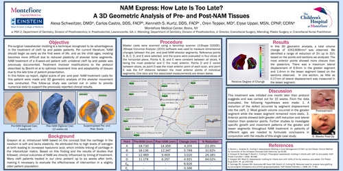
Procedure: Master casts were scanned using a benchtop scanner (3-Shape D2000). 3Shape Convince Analyzer (2015) software was used to measure dimensional changes between the pre- and post-NAM alveolar segments. Reference points A, B, C, D, and E were selected, and the scans were evaluated in four slices of the horizontal plane. Points A, B, and C were constant between all slices, A being the most posterior and C the most anterior. Points D and E varied between slices, as point D was the most anterior point of each scan, and point E was the A-P distance between the most anterior points of individual segments. One slice and the associated measurements are shown below.
Results: In this 3D geometric analysis, a total volume change of 1043.898mm3 was observed. We identified a range of reduction of 3% up to 31% based on the points and sections we analyzed. The most anterior points showed more closure than the posteriors. There was a maximum lateral displacement of 6.9mm in the greater segment and 1.4mm in the lesser segment based on the sections observed. In one section, as little as 0.17mm of lateral displacement was measured in the lesser segment.
Discussion: This treatment was initiated one month later than protocol suggests and was carried out for 15 weeks. From the data evaluated, the following hypotheses were made: 1. A reduction of the defect occurred by segment displacement into the cleft. 2. Most growth volume occurred in the greater segment while the lesser segment remained more static. 3. Anterior points showed both greater cleft reduction and lateral rotation than posterior points. Further studies to investigate specific growth and movement patterns of the greater and lesser segments throughout NAM treatment in patients of different ages are needed to formulate conclusions in conjunction with the results of this single case study.
References:
1. Brecht L., Grayson B., Cutting C. Nasoalveolar Molding in Early Management of Cleft Lip and Palate. Clinical Method for Correction of the Unilateral Oronasal Cleft Deformity. pp 63-84.
2. Grayson B, Santiago P, Brecht L, Cutting C. Presurgical nasoalveolar molding in infants with cleft lip and palate. Cleft Palate Craniofac J 1999; 36:486.
3. Grayson BH, Maull D. Nasoalveolar molding for infants born with clefts of the lip, alveolus, and palate. Clin Plastic Surg 2004; 34. pp 149-158.
4. Santiago PE, Grayson BH, Gianoutos MP, Kwon SM, Brecht LE, Cutting CB. Reduced need for alveolar bone grafting by pre-surgical orthopedics and primary gingivoperioplasty. Cleft Palate Craniofac J. 1998; 35: 77-80.