66 posters, 2 videos, 1 audios, 2 topics, 162 authors, 49 institutions
ePostersLive by SciGen Technologies S.A. All rights reserved.
American College of Prosthodontists - 2020 ANNUAL SESSION
November 4-7 | Virtual Meeting

55
Application of Intra-Oral Scanner at the Implant placement for the Modified "One Abutment One Time" Concept
Poster Presenter
Authors
Yoshiatsu Tanaka, Chao-Chieh Yang, Yusuke Hamada, Wei-Shao Lin
Affiliations
Part of Topic
Case Presentation
Application of Intra-Oral Scanner at the Implant placement for the Modified "One Abutment One Time" Concept
The “One Abutment One Time” concept was introduced to reduce the soft tissue disturbance upon abutment disconnections and reconnections (Cocchetto 2010, Canullo 2010, Degidi 2011).
However, a final abutment placement cannot be achieved when the site requires primary closure.
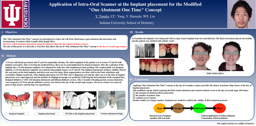
The aim of this poster is to describe a work flow that allows the use of “One Abutment One Time” concept at the day of second stage surgery
A 19-year-old female presented with #7 and 10 congenitally missing. The chief complaint of the patient was to restore #7 and 10 with implant restoration. After reviewing the medical history, there are no contraindication for dental treatment. After the evaluation of the implant sites, 2.9 mm diameter implants were planned for both sites with simultaneous bone grafting. The surgical guide was designed digitally. A 2.9 mm diameter implants were placed using guided surgery on #7 and 10. The implants had proper primary stability. Placed the scan body on the both implants and intra-oral scan was taken. Bone augmentation was done with bovine bone substitutes and resorbable collagen membrane. After implant placement, two STL files (one is diagnostic cast and the other one is at the time of implant placement) were superimposed, and the position of subgingival margin was predicted. Following the determination of the marginal line, designed definitive CAD/CAM titanium abutments and lithium disilicate crowns. After 2 months of healing period, custom abutments were torqued to 35 N/cm and the definitive crowns were luted at the day of the second stage surgery. All excess cement was removed prior to flap closure, and the flap was repositioned.
At 8 months the implants were integrated with no signs of peri-implant bone loss and infection. The final restorations placed successfully and the patient was satisfied with esthetic result.
Applying “One Abutment One Time” concept at the day of secondary surgery provides the chance of primary flap closure at the day of implant placement.
This modified concept which is placing the final custom abutment and cement-retained crown at the day of second stage will reduce
① the amounts of abutment dis/reconnections
② the number of patient visits
③ residual excess cement.