203 posters, 14 topics, 5 sessions, 817 authors, 256 institutions
ePostersLive by SciGen Technologies S.A. All rights reserved.
The AAPS 102nd Annual Meeting
May 18-21, 2024 | The Westin Copley Place, Boston, MA

D19
Accuracy Of Preoperative Imaging In Assessing The Involvement Of The Deep Periosteal Margin In Dermatofibrosarcoma Protuberans Of The Scalp: The Mayo Clinic Experience
Poster Presenter
Authors
Sai Cherukuri, Eugene Zheng, Jerry Brewer, Julie Guerin, Uldis Bite, Samir Mardini, Waleed Gibreel
Affiliations
Mayo Clinic Rochester, Division of Plastic and Reconstructive Surgery, Mayo Clinic Rochester, Department of Dermatology, Mayo Clinic Rochester, Department of Radiology, Mayo Clinic Rochester
Part of Topic
Craniofacial/Peds
Accuracy of Preoperative Imaging in Assessing the involvement of the Deep Periosteal Margin in Dermatofibrosarcoma Protuberans of the Scalp: The Mayo Clinic Experience
Background
Dermatofibrosarcoma protuberans (DFSP) of the scalp may infiltrate the periosteum, calvarium, dura or brain. Preoperative imaging (CT and/or MRI) can show calvarial bone abnormalities, but it remains to be seen whether these abnormalities correlate with the positive deep periosteal margins at excision.
Objective
We sought to examine the accuracy of preoperative CT/MRI in assessment of the deep margin and whether preoperative calvarial bone abnormalities correlate with the involvement of the deep periosteal margin.
Methods
A retrospective medical record and image review was done of all patients with DFSP of the scalp treated at a quaternary referral center. Data collected included demographic, gross tumor characteristics, preoperative imaging findings, and status of the deep periosteal margin (involved versus non-involved).
Results
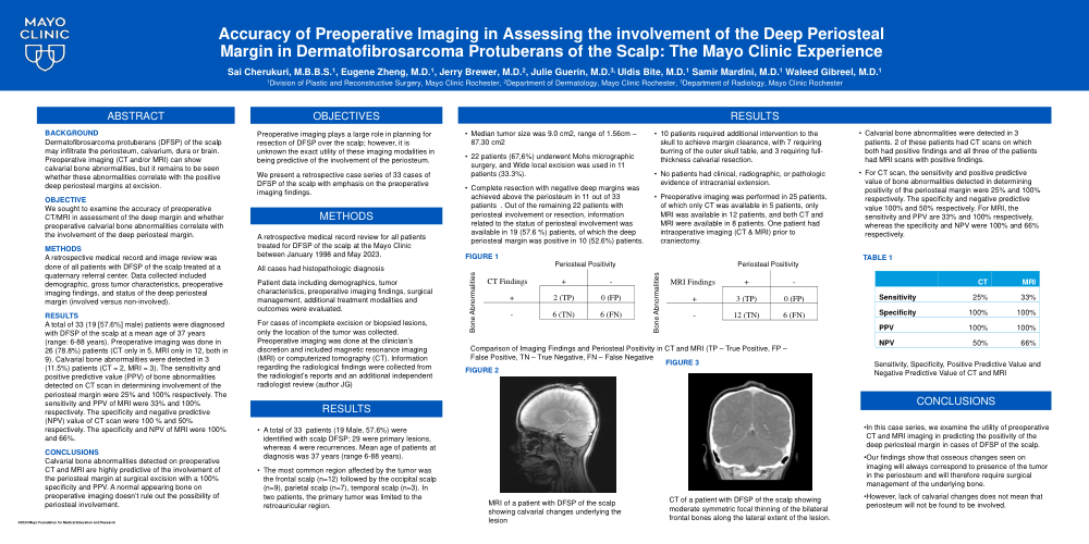
A total of 33 (19 [57.6%] male) patients were diagnosed with DFSP of the scalp at a mean age of 37 years (range: 6-88 years). Preoperative imaging was done in 26 (78.8%) patients (CT only in 5, MRI only in 12, both in 9). Calvarial bone abnormalities were detected in 3 (11.5%) patients (CT = 2, MRI = 3). The sensitivity and positive predictive value (PPV) of bone abnormalities detected on CT scan in determining involvement of the periosteal margin were 25% and 100% respectively. The sensitivity and PPV of MRI were 33% and 100% respectively. The specificity and negative predictive (NPV) value of CT scan were 100 % and 50% respectively. The specificity and NPV of MRI were 100% and 66%.
Conclusions
Calvarial bone abnormalities detected on preoperative CT and MRI are highly predictive of the involvement of the periosteal margin at surgical excision with a 100% specificity and PPV. A normal appearing bone on preoperative imaging doesn’t rule out the possibility of periosteal involvement.