203 posters, 14 topics, 5 sessions, 817 authors, 256 institutions
ePostersLive by SciGen Technologies S.A. All rights reserved.
The AAPS 102nd Annual Meeting
May 18-21, 2024 | The Westin Copley Place, Boston, MA

D160
Rates Of Dehiscence And Fistula Formation Following Anterior Palatal Reconstruction With Vomer Flap
Poster Presenter
Authors
Jesse Menville, Carole Spake, Luke Soliman, Nidhi Shinde, Elijah Persad-Paisley, Navya Baranwal, Albert Woo
Affiliations
Part of Topic
Craniofacial/Peds
INTRODUCTION
The Role of the Vomer Bone
Vomer flap reconstruction is a surgical procedure used to repair a unilateral or bilateral cleft palate. In this procedure, a flap of vomer mucoperiosteal tissue is mobilized and repositioned to cover the cleft. One side of the tissue remains attached to the bone to retain its original blood supply, while the other is sutured to the lateral nasal or oral wall.
Advantages
Disadvantages
METHODS
A retrospective review was conducted of all cleft palate repairs conducted by the senior author over a seven-year period. All patients who underwent vomer flap reconstruction prior to palatoplasty were assigned to the vomer flap cohort. Those who underwent palatoplasty alone were assigned to the control cohort.
Primary outcomes included dehiscence after vomer flap reconstruction (vomer flap cohort only) and fistula formation of the anterior hard palate (both cohorts). Fistula formation was assessed no sooner than three months and no later than one year after palatoplasty. All vomer flap reconstructions were performed using a superior-based flap technique.
RESULTS
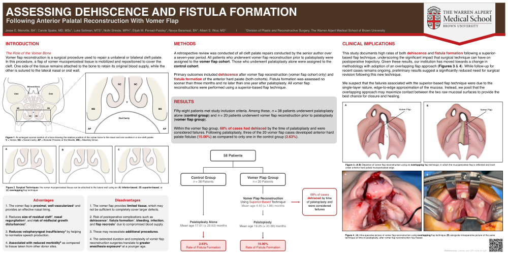
Fifty-eight patients met study inclusion criteria. Among these, n = 38 patients underwent palatoplasty alone (control group) and n = 20 patients underwent vomer flap reconstruction prior to palatoplasty (vomer flap group).
Within the vomer flap group, 68% of cases had dehisced by the time of palatoplasty and were considered failures. Following palatoplasty, three of the 20 vomer flap cases developed anterior hard palate fistulas (15.00%) as compared to only one in the control group (2.63%).
CLINICAL IMPLICATIONS
This study documents high rates of both dehiscence and fistula formation following a superior-based flap technique, underscoring the significant impact that surgical technique can have on postoperative trajectory. Given these results, our institution has moved towards a change in methodology with adoption of an overlapping flap approach (Figures 3 & 4). While follow-up for recent cases remains ongoing, preliminary results suggest a significantly reduced need for surgical revision following this new technique.
We suspect that the failures associated with the superior-based flap technique were due to the single-layer nature, edge-to-edge approximation of the mucosa. Instead, we posit that the overlapping approach may maximize contact between the two raw mucosal surfaces to provide the best chance for closure and healing.