4,068 posters, 219 videos, 101 topics, 69 sessions, 14,365 authors, 6,128 institutions
ePostersLive by SciGen Technologies S.A. All rights reserved.
Alzheimer's Association International Conference 2026
July 12-15, 2026 | London, United Kingdom, and Online

13721
Bone-brain Axis In Alzheimer’S Disease: Associations Among Bone Mineral Density, Inflammation, Tau Pathology And Gray Matter Atrophy
Part of Topic
Virtual
Bone-brain axis in Alzheimer’s disease: associations among bone mineral density, inflammation, tau pathology and gray matter atrophy
Fan Zhang1, Jing Li1,2, Tenghong Lian1,2, Jinghui Li1, Dongmei Luo1, Lu Rong1, Zhan Liu1, Yao Meng1, Yingdi Liu1,Wei Zhang 1,2*
1 Department of Neurology, Beijing Tiantan Hospital, Capital Medical University, Beijing,100070, China.
2 Center for Cognitive Neurology, Department of Neurology, Beijing Tiantan Hospital, Capital Medical University, Beijing,100070, China.
First author: Fan Zhang
*Correspondence to: Wei Zhang
Full address: Beijing Tiantan Hospital, 119 South Fourth Ring Road West, Fengtai District, Beijing, China
Email: ttyyzw@163.com
Abstract
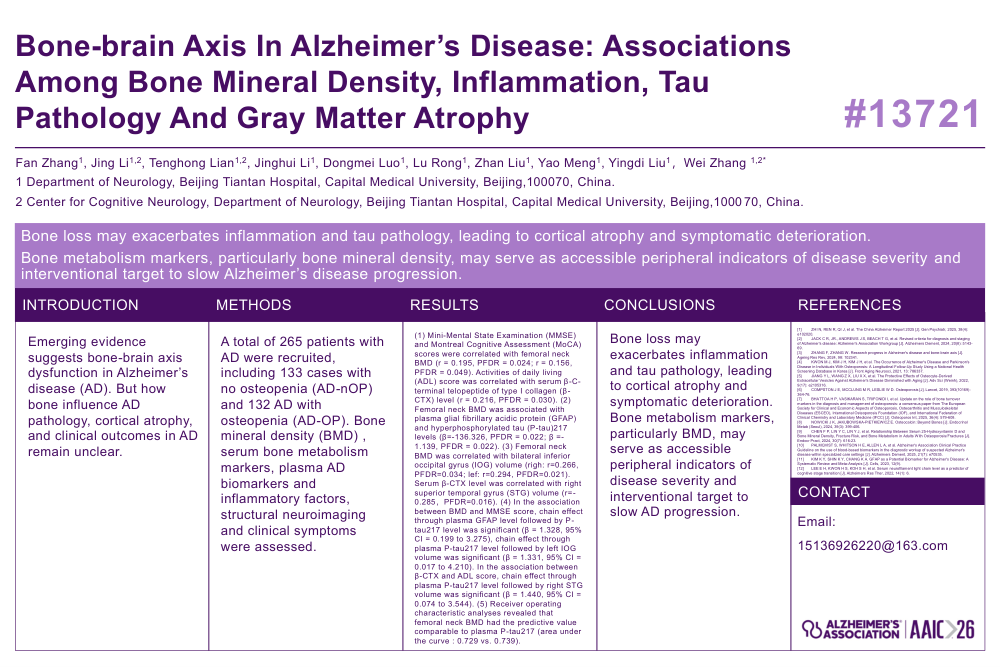
Background: Emerging evidence suggests bone-brain axis dysfunction in Alzheimer’s disease (AD). But how bone influence AD pathology, cortical atrophy, and clinical outcomes in AD remain unclear.
Methods: A total of 265 patients with AD were recruited, including 133 cases with no osteopenia (AD-nOP) and 132 AD with osteopenia (AD-OP). Bone mineral density (BMD), serum bone metabolism markers, plasma AD biomarkers and inflammatory factors, structural neuroimaging and clinical symptoms were assessed.
Results: (1) Mini-Mental State Examination (MMSE) and Montreal Cognitive Assessment (MoCA) scores were correlated with femoral neck BMD (r = 0.195, PFDR = 0.024; r = 0.156, PFDR = 0.049). Activities of daily living (ADL) score was correlated with serum β-C-terminal telopeptide of type I collagen (β-CTX) level (r = 0.216, PFDR = 0.030). (2) Femoral neck BMD was associated with plasma glial fibrillary acidic protein (GFAP) and hyperphosphorylated tau (P-tau)217 levels (β=-136.326, PFDR = 0.022; β =-1.139, PFDR = 0.022). (3) Femoral neck BMD was correlated with bilateral inferior occipital gyrus (IOG) volume (righ: r=0.266, PFDR=0.034; lef: r=0.294, PFDR=0.021). Serum β-CTX level was correlated with right superior temporal gyrus (STG) volume (r=-0.285,PFDR=0.016). (4) In the association between BMD and MMSE score, chain effect through plasma GFAP level followed by P-tau217 level was significant (β = 1.328, 95% CI = 0.199 to 3.275), chain effect through plasma P-tau217 level followed by left IOG volume was significant (β = 1.331, 95% CI = 0.017 to 4.210). In the association between β-CTX and ADL score, chain effect through plasma P-tau217 level followed by right STG volume was significant (β = 1.440, 95% CI = 0.074 to 3.544). (5) Receiver operating characteristic analyses revealed that femoral neck BMD had the predictive value comparable to plasma P-tau217 (area under the curve : 0.729 vs. 0.739).
Conclusion: Bone loss may exacerbates inflammation and tau pathology, leading to cortical atrophy and symptomatic deterioration. Bone metabolism markers, particularly BMD, may serve as accessible peripheral indicators of disease severity and interventional target to slow AD progression.
【Keywords】Alzheimer’s disease; bone-brain axis; bone mineral density; β-C-terminal telopeptide of type I collagen; glial fibrillary acidic protein; hyperphosphorylated tau 217; cortical atrophy; clinical symptoms.
Comments
Email is required but is not shown publicly.
Loading comments...