4,068 posters, 219 videos, 101 topics, 69 sessions, 14,365 authors, 6,128 institutions
ePostersLive by SciGen Technologies S.A. All rights reserved.
Alzheimer's Association International Conference 2026
July 12-15, 2026 | London, United Kingdom, and Online

13264
Retinal Microvascular Changes During Donanemab Therapy In Early Alzheimer’S Disease: A 6-month Longitudinal Study.
Part of Topic
Virtual
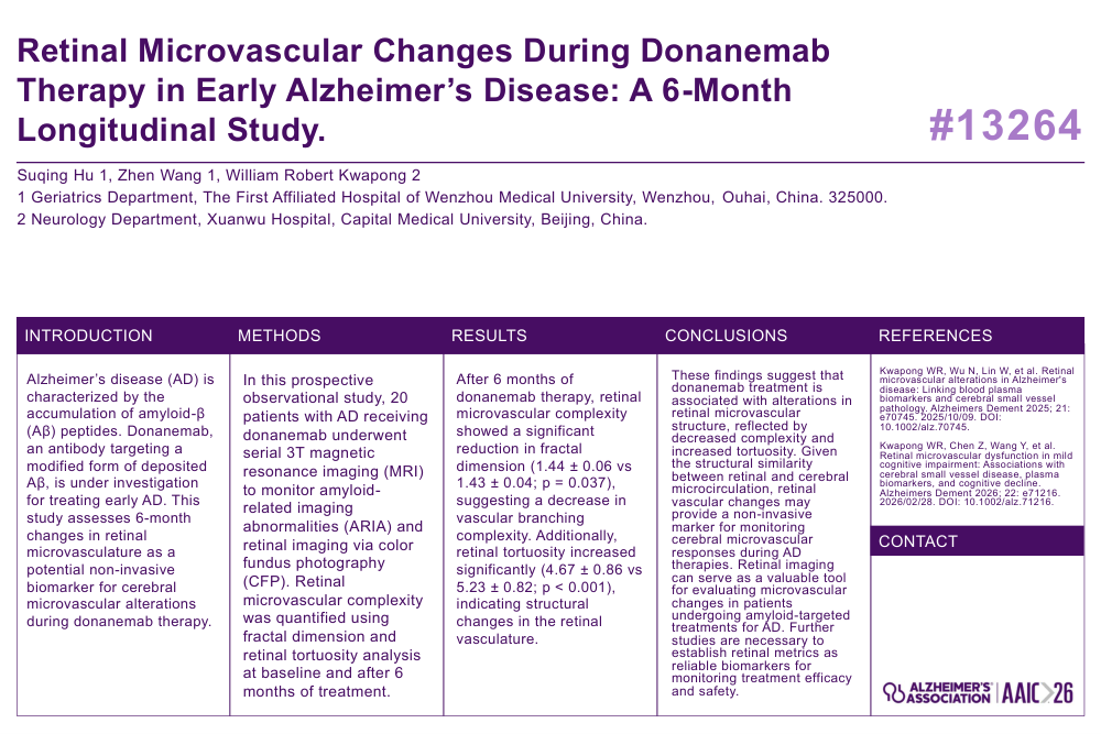
Retinal Microvascular Changes During Donanemab Therapy in Early Alzheimer’s Disease: A 6-Month Longitudinal Study.
Suqing Hu 1, Zhen Wang 1, William Robert Kwapong 2
1 Geriatrics Department, The First Affiliated Hospital of Wenzhou Medical University, Wenzhou, Ouhai, China. 325000.
2 Neurology Department, Xuanwu Hospital, Capital Medical University, Beijing, China.
INTRODUCTION
Alzheimer’s disease (AD) is characterized by the accumulation of amyloid-β (Aβ) peptides. Donanemab, an antibody targeting a modified form of deposited Aβ, is under investigation for treating early AD. This study assesses 6-month changes in retinal microvasculature as a potential non-invasive biomarker for cerebral microvascular alterations during donanemab therapy
METHODS
In this prospective observational study, 20 patients with AD receiving donanemab underwent serial 3T magnetic resonance imaging (MRI) to monitor amyloid-related imaging abnormalities (ARIA) and retinal imaging via color fundus photography (CFP). Retinal microvascular complexity was quantified using fractal dimension and retinal tortuosity analysis at baseline and after 6 months of treatment.
RESULTS
After 6 months of donanemab therapy, retinal microvascular complexity showed a significant reduction in fractal dimension (1.44 ± 0.06 vs 1.43 ± 0.04; p = 0.037), suggesting a decrease in vascular branching complexity. Additionally, retinal tortuosity increased significantly (4.67 ± 0.86 vs 5.23 ± 0.82; p < 0.001), indicating structural changes in the retinal vasculature.
CONCLUSIONS
These findings suggest that donanemab treatment is associated with alterations in retinal microvascular structure, reflected by decreased complexity and increased tortuosity. Given the structural similarity between retinal and cerebral microcirculation, retinal vascular changes may provide a non-invasive marker for monitoring cerebral microvascular responses during AD therapies. Retinal imaging can serve as a valuable tool for evaluating microvascular changes in patients undergoing amyloid-targeted treatments for AD. Further studies are necessary to establish retinal metrics as reliable biomarkers for monitoring treatment efficacy and safety.
REFERENCES
Kwapong WR, Wu N, Lin W, et al. Retinal microvascular alterations in Alzheimer's disease: Linking blood plasma biomarkers and cerebral small vessel pathology. Alzheimers Dement 2025; 21: e70745. 2025/10/09. DOI: 10.1002/alz.70745.
Kwapong WR, Chen Z, Wang Y, et al. Retinal microvascular dysfunction in mild cognitive impairment: Associations with cerebral small vessel disease, plasma biomarkers, and cognitive decline. Alzheimers Dement 2026; 22: e71216. 2026/02/28. DOI: 10.1002/alz.71216.
Comments
Email is required but is not shown publicly.
Loading comments...