4,068 posters, 219 videos, 101 topics, 69 sessions, 14,365 authors, 6,128 institutions
ePostersLive by SciGen Technologies S.A. All rights reserved.
Alzheimer's Association International Conference 2026
July 12-15, 2026 | London, United Kingdom, and Online

13377
Automated Ct-based Brain Volumetry For Resource-limited Settings
Part of Topic
Virtual
Automated CT-Based Brain Volumetry for Resource-Limted Settings
Background:
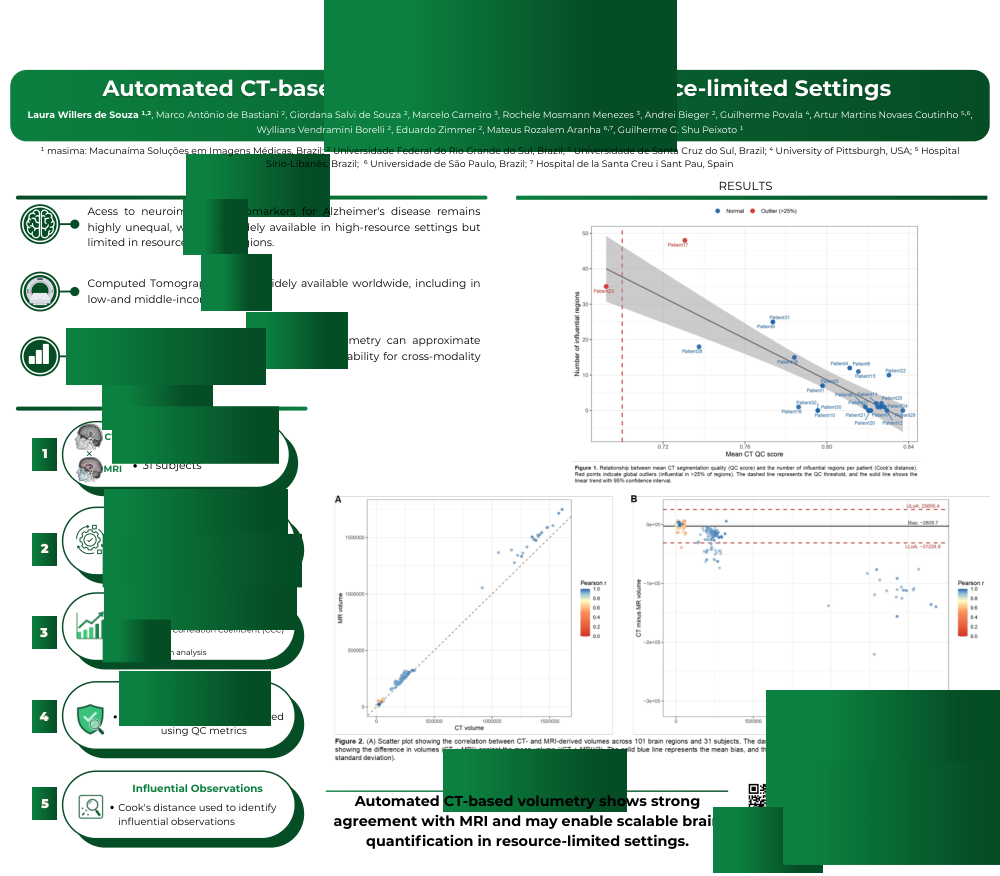
Access to neuroimaging biomarkers for Alzheimer’s disease remains highly unequal, with Magnetic Resonance Imaging (MRI)-based volumetry mostly restricted to high-resource settings. In contrast, Computed Tomography (CT) is widely available worldwide. Enabling reliable quantitative analysis from CT scans could reduce disparities in dementia care. This study evaluates whether automated CT-based volumetry can approximate MRI-derived measurements with sufficient reliability for cross-modality comparability.
Methods:
Paired CT and MRI scans from 31 subjects were analyzed using deep learning–based automated segmentation pipelines to extract regional brain volumes. Cross-modality agreement was assessed using Pearson correlation, Lin’s concordance correlation coefficient (CCC), intraclass correlation coefficient (ICC[2,1]) for absolute agreement, and Bland–Altman analysis. Segmentation quality control (QC) metrics were used to assess the impact of quality, and Cook’s distance identified influential observations.
Results:
Automated CT-based volumetry showed strong correlation with MRI (mean Pearson r = 0.79), with 100/101 regions showing significant correlation after FDR correction (Figure 1A). Agreement was region-dependent: 11 regions showed excellent reliability (ICC > 0.9) and 29 good (0.75–0.9), while 18 regions—including cerebellar and selected limbic and occipital regions—demonstrated poor agreement (ICC < 0.5). CCC (mean = 0.67) indicated reduced concordance due to systematic bias. Bland–Altman analysis revealed consistent underestimation of CT-derived volumes (−2,661 mm³) (Figure 1B), further reflected by CT/MRI ratios below unity across regions (Figure 2). Lower CT QC scores were associated with increased statistical influence and reduced agreement, highlighting image quality as a key determinant of real-world reliability (Figure 3).
Conclusions:
Automated CT-based volumetry captures clinically relevant structural information with strong correlation to MRI, supporting its role as a scalable alternative for brain quantification. While not fully interchangeable, its performance across most brain regions enables population-level screening and longitudinal monitoring where MRI is limited. Using routine CT scans, this approach provides a cost-effective pathway to expand access to neuroimaging biomarkers in underserved settings. Although modality-specific calibration remains necessary, it represents a critical step toward more equitable access to dementia diagnosis and monitoring.
Keywords:
Brain volumetry; Computed tomography; Magnetic resonance imaging; Alzheimer's disease; Dementia; Brain atrophy; Neuroimaging biomarkers; Quantitative imaging; Deep learning; Resource-limited settings.
Comments
Email is required but is not shown publicly.
Loading comments...