4,068 posters, 219 videos, 101 topics, 69 sessions, 14,365 authors, 6,128 institutions
ePostersLive by SciGen Technologies S.A. All rights reserved.
Alzheimer's Association International Conference 2026
July 12-15, 2026 | London, United Kingdom, and Online

12950
AI-derived Vascular Age From Photoplethysmography Is Associated With Brain Structural Alterations Across Major Brain Networks
Poster Presenter
Authors
Jiong Chen, Guangkun Nie, Xiaqing Jiang, Yaqing Gao, Junhao Wen, Song Gao, Shenda Hong, Yue Leng
Affiliations
Department of Psychiatry and Behavioral Sciences, University of California, San Francisco, CA, USA, School of Intelligence Science and Technology, Peking University, Beijing, China, Department of Population Health, New York University Grossman School of Medicine, New York, NY, USA, Department of Epidemiology and Public Health, Institute of Epidemiology and Health Care, University College London, London, UK, Laboratory of AI and Biomedical Science (LABS), Columbia University, New York, NY, USA, Institute of Medical Technology, Peking University Health Science Center, Beijing, China, National Institute of Health Data Science, Peking University, Beijing, China
Part of Topic
Virtual
Background:
Vascular aging is a key contributor to dementia, yet traditional cardiovascular risk factors do not fully capture vascular health. Vascular age, estimated from photoplethysmography (PPG) signals using deep learning, provides a non-invasive and scalable biomarker of vascular aging. However, its associations with brain structure remain unclear.
Methods:
We applied a validated deep learning model to derive vascular age from PPG recordings in 14,593 UK Biobank participants. Vascular age gap (VAG) was defined as the difference between PPG-predicted and chronological age. Multivariable linear regression models were used to examine associations between VAG and multimodal magnetic resonance imaging (MRI) markers, adjusting for sociodemographic factors, lifestyles, APOE ε4 status, baseline cardiovascular risk factors, time from PPG to MRI, and imaging-related confounders. Sensitivity analyses further accounted for follow-up cardiovascular risk factors and excluded incident cardiovascular diseases (CVD). All p values were corrected for multiple comparisons.
Results:
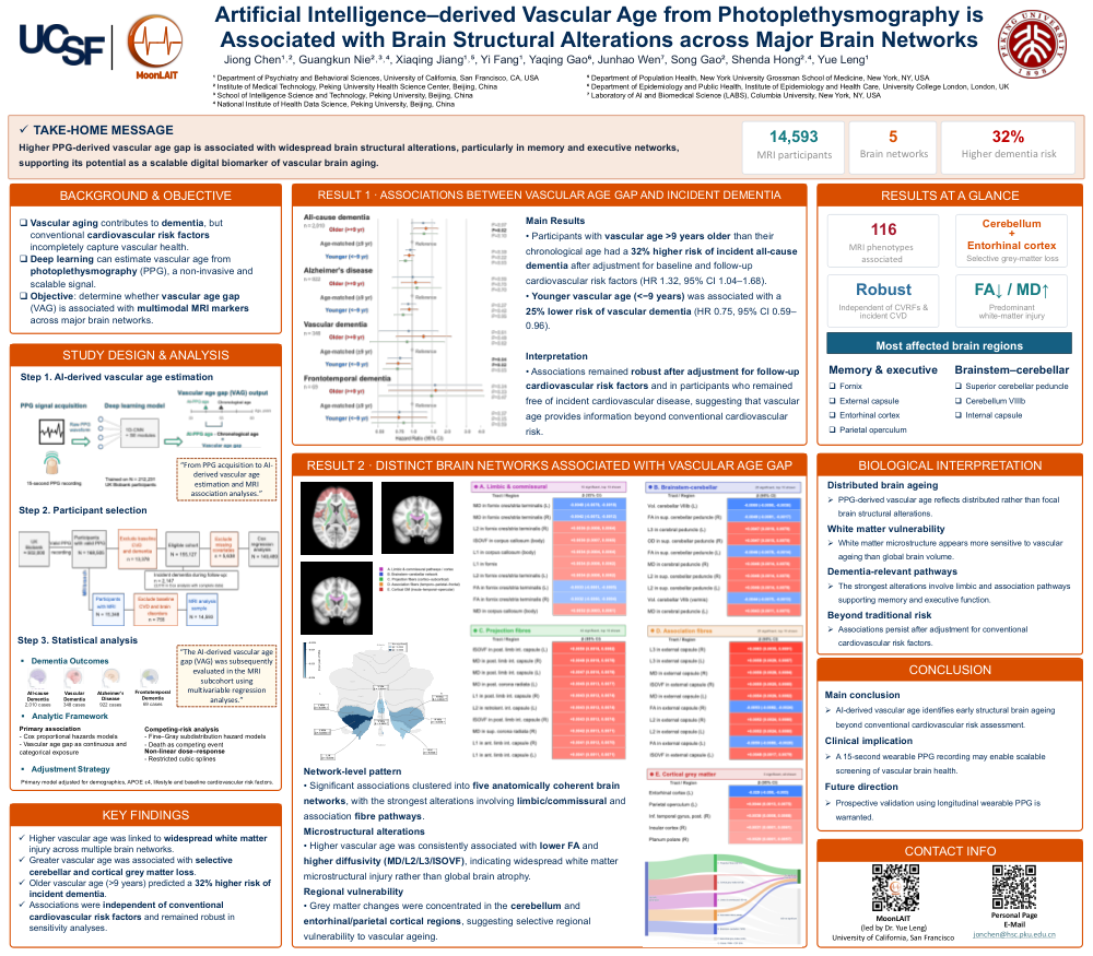
Higher VAG was associated with widespread structural adverse alterations across major brain networks (Fig. 1). In dementia-relevant regions, greater VAG was linked to microstructural damage in memory-related limbic circuits (e.g., fornix: fractional anisotropy [FA] β = -0.0029, p = 0.041; mean diffusivity [MD] β = 0.0031, p = 0.027) and executive function pathways (e.g., external capsule: FA β = -0.0053, p < 0.001). In addition, cortical grey matter analysis showed reduced volumes in the parietal operculum and cerebellum, regions underlying spatial and sensory integration. By contrast, associations in non-cognitive pathways were more localized, including projection fibers (internal capsule MD β = 0.0048, p = 0.002) and brainstem–cerebellar pathways (superior cerebellar peduncle FA β = -0.0049, p = 0.003) (Fig. 2). These findings were consistent in sensitivity analyses.
Conclusions:
PPG-derived vascular age captures distinct patterns of structural brain aging, particularly within memory and executive networks. These findings support its potential as a scalable digital biomarker for detecting vascular contributions to neurodegeneration.
Comments
Email is required but is not shown publicly.
Loading comments...