4,068 posters, 219 videos, 101 topics, 69 sessions, 14,365 authors, 6,128 institutions
ePostersLive by SciGen Technologies S.A. All rights reserved.
Alzheimer's Association International Conference 2026
July 12-15, 2026 | London, United Kingdom, and Online

13626
A Multimodal Approach To Characterizing Molecular Interactions Between Traumatic Brain Injury And Alzheimer’S Disease
Poster Presenter
Authors
Part of Topic
Virtual
A multi-modal approach to characterizing molecular interactions between traumatic brain injury and Alzheimer’s disease
Mehwish Anwer1,2, Mckenna Stuart1,2, Larissa Kraus3, Shalini Iyer3, Wai Hang Cheng1,2, Jianjia Fan1,2, Tetiana Poliakova1,2, Anna Wilkinson1,2, Carlos Barron1,2, Mark S. Cembrowski*1,3,Cheryl L. Wellington*1,2, *co-senior authors
1Djavad Mowafaghian Centre for Brain Health, University of British Columbia, Vancouver, Canada
2Department of Pathology and Laboratory Medicine, University of British Columbia, Vancouver, Canada
3Department of Cellular and Physiological Sciences, Life Sciences Institute, University of British Columbia, Vancouver, Canada
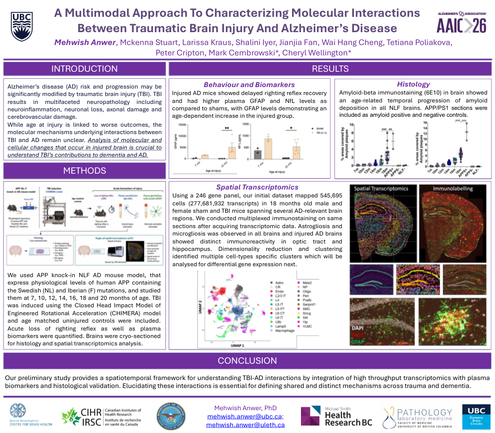
Background: Alzheimer’s disease (AD) risk and progression may be significantly modified by traumatic brain injury (TBI), an important yet underexplored factor in neurodegeneration. While age at injury is linked to worse outcomes, the molecular mechanisms underlying interactions between TBI and AD remain unclear. Therefore, we investigated molecular targets that are acutely dysregulated upon TBI in murine brain with or without amyloid pathology using spatial transcriptomics approach combined with biomarker and histopathological analysis.
Methods: We used APP knock-in NLF AD mouse model that express physiological levels of human APP containing the Swedish (NL) and Iberian (F) mutations and studied them at 7, 10, 12, 14, 16, 18 and 20 months of age. TBI was induced using the Closed Head Impact Model of Engineered Rotational Acceleration (CHIMERA) model and age matched uninjured controls were included. Acute loss of righting reflex (LRR) was assessed. Acute plasma neurofilament light (NfL) and glial fibrillary acidic protein (GFAP) were quantified as biomarkers of axonal injury and astrocyte activation, respectively. Brains were cryosectioned for histology and a subset was prepared for single cell spatial transcriptomics analysis (scST; Xenium by 10X Genomics).
Results: Injured mice showed delayed righting reflex recovery. NLF mice exhibit acute increases in plasma GFAP and NfL levels following CHIMERA injury, with GFAP levels demonstrating an age-dependent increase in the injured group. Amyloid-beta immunostaining in brain showed an age-related temporal progression of amyloid deposition in all NLF brains whereas distinct astrogliosis and microgliosis was observed in optic tract and hippocampus of injured brains. Using a 246 gene panel, our initial scST dataset mapped 545,695 cells (277,681,932 transcripts) in 18 months old mice (TBI=4, sham=3) spanning several AD-relevant brain regions. Dimensionality reduction and clustering identified multiple dysregulated genes in clusters corresponding to major cell types including neurons, astrocytes, endothelial cell, microglia, oligodendrocyte, pericyte and vascular leptomeningeal cells.
Conclusion: The study provides a spatiotemporal framework for understanding TBI-AD interactions through integration of high throughput transcriptomics with plasma biomarkers and histological validation. Elucidating these interactions is essential to define shared and distinct mechanisms across aging, trauma and dementia.
Comments
Email is required but is not shown publicly.
Loading comments...