193 posters, 19 videos, 10 audios, 3 topics, 28 sessions, 714 authors, 278 institutions
ePostersLive by SciGen Technologies S.A. All rights reserved.
9th World Congress on Vascular Access 2026
15 - 17 April, 2026 | Valencia, Spain

36
Vascular Access in the morbidly obese: use of a spinal needle, a case report and review
Poster Presenter
Authors
Affiliations
Part of Topic
Introduction: The use of spinal needles(SN) for vascular access(VA) is rarely mentioned in the literature, and no detailed description of its use in conjunction with US for placement of an axillary CICC has been reported.
Methods: We present a case in which a SN was used to place an axillary CICC under US in an obese patient with limited options. This is, to the best of our knowledge, the first reported case of this technique. We also present a literature review of the use of SN for VA.
Case: A 40 yo morbidly obese (BMI 62,8) male patient suffered a high energy motor vehicle collision, EMS was unable to obtain VA, in the ED a Short Peripheral Catheter (SPC) was secured and after a CT he was taken to the OR, where 2 more SPCs were secured after 1 failed attempt to place a CVC, later in the ICU the attending physician failed to obtain a (blind) subclavian access due to vein depth, so a 4 lumens right inguinal FICC was placed under US. 24 hours later we where asked to help establish a better and safer access due to concerns for the risk of infection.
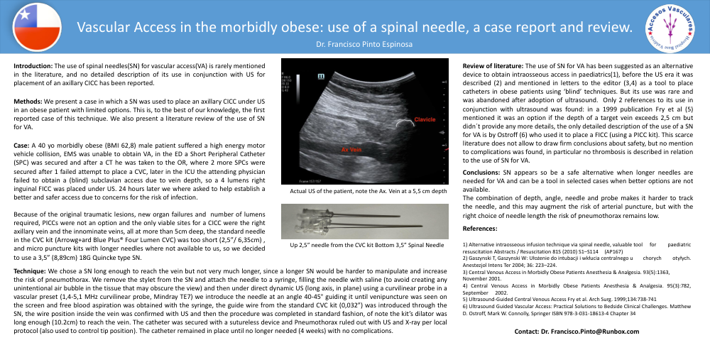
Because of the original traumatic lesions, new organ failures and number of lumens required, PICCs were not an option and the only viable sites for a CICC were the right axillary vein and the innominate veins, all at more than 5cm deep, the standard needle in the CVC kit (Arrowg+ard Blue Plus® Four Lumen CVC) was too short (2,5”/ 6,35cm) , and micro puncture kits with longer needles where not available to us, so we decided to use a 3,5” (8,89cm) 18G Quincke type SN.
Technique: We chose a SN long enough to reach the vein but not very much longer, since a longer SN would be harder to manipulate and increase the risk of pneumothorax. We remove the stylet from the SN and attach the needle to a syringe, filling the needle with saline (to avoid creating any unintentional air bubble in the tissue that may obscure the view) and then under direct dynamic US (long axis, in plane) using a curvilinear probe in a vascular preset (1,4-5,1 MHz curvilinear probe, Mindray TE7) we introduce the needle at an angle 40-45° guiding it until venipuncture was seen on the screen and free blood aspiration was obtained with the syringe, the guide wire from the standard CVC kit (0,032”) was introduced through the SN, the wire position inside the vein was confirmed with US and then the procedure was completed in standard fashion, of note the kit’s dilator was long enough (10.2cm) to reach the vein. The catheter was secured with a sutureless device and Pneumothorax ruled out with US and X-ray per local protocol (also used to control tip position). The catheter remained in place until no longer needed (4 weeks) with no complications.
Review of literature: The use of SN for VA has been suggested as an alternative device to obtain intraosseous access in paediatrics(1), before the US era it was described (2) and mentioned in letters to the editor (3,4) as a tool to place catheters in obese patients using ‘blind’ techniques. But its use was rare and was abandoned after adoption of ultrasound. Only 2 references to its use in conjunction with ultrasound was found: in a 1999 publication Fry et al (5) mentioned it was an option if the depth of a target vein exceeds 2,5 cm but didn´t provide any more details, the only detailed description of the use of a SN for VA is by Ostroff (6) who used it to place a FICC (using a PICC kit). This scarce literature does not allow to draw firm conclusions about safety, but no mention to complications was found, in particular no thrombosis is described in relation to the use of SN for VA.
Conclusions: SN appears so be a safe alternative when longer needles are needed for VA and can be a tool in selected cases when better options are not available. The combination of depth, angle, needle and probe makes it harder to track the needle, and this may augment the risk of arterial puncture, but with the right choice of needle length the risk of pneumothorax remains low.
References: 1) Alternative intraosseous infusion technique via spinal needle, valuable tool for paediatric resuscitation Abstracts / Resuscitation 81S (2010) S1–S114 (AP167) 2) Gaszynski T, Gaszynski W: Ułożenie do intubacji i wkłucia centralnego u chorych otyłych. Anestezjol Intens Ter 2004; 36: 223–224. 3) Central Venous Access in Morbidly Obese Patients Anesthesia & Analgesia. 93(5):1363, November 2001. 4) Central Venous Access in Morbidly Obese Patients Anesthesia & Analgesia. 95(3):782, September 2002. 5) Ultrasound-Guided Central Venous Access Fry et al. Arch Surg. 1999;134:738-741 6) Ultrasound Guided Vascular Access: Practical Solutions to Bedside Clinical Challenges. Matthew D. Ostroff, Mark W. Connolly, Springer ISBN 978-3-031-18613-4 Chapter 34
Contact: Dr. Francisco.Pinto@Runbox.com