193 posters, 19 videos, 10 audios, 3 topics, 28 sessions, 709 authors, 279 institutions
ePostersLive by SciGen Technologies S.A. All rights reserved.
9th World Congress on Vascular Access 2026
15 - 17 April, 2026 | Valencia, Spain

166
<p>Comparison of Vein Selection of a Novel Ultrasound-Based Vein Visualization Device and Traditional Landmark Technique</p>
Poster Presenter
Authors
Michael Liddelow, Gavin Jackson, Kate Molan, Ellaby Hansen, Peter Carr, Barry Doyle, Nikhilesh Bappoo
Affiliations
UWA Centre for Medical Research (affiliated with the Harry Perkins Institute of Medical Research), School of Nursing and Midwifery, University of Galway, School of Physics, Mathematics and Computing, The University of Western Australia, Fiona Stanley Hospital, Western Australia, VeinTech Pty Ltd, Western Australia, Vascular Engineering Laboratory, Harry Perkins Institute of Medical Research, QEII Medical Centre Nedlands and Centre for Medical Research, The University of Western Australia
Part of Topic
Comparison of Vein Selection of a Novel Ultrasound-Based Vein Visualization Device and Traditional Landmark Technique
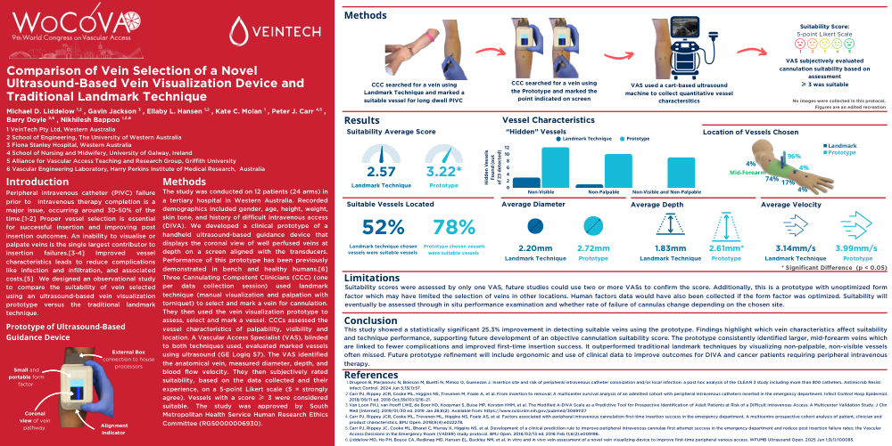
Peripheral intravenous catheter (PIVC) failure prior tointravenous therapy completion is a major issue, occurring around 30-50% of the time.[1-2] Proper vessel selection is essential for successful insertion and improving post insertion outcomes. An inability to visualise or palpate veins is the single largest contributor to insertion failures.[3-4] Improved vessel characteristics leads to reduce complications like infection and infiltration, and associated costs.[5]We designed an observational study to compare the suitability of vein selected using an ultrasound-based vein visualization prototype versus the traditional landmark technique.
The study was conducted on 12 patients (24 arms) in a tertiary hospital in Western Australia. Recorded demographics included gender, age, height, weight, skin tone, and history of difficult intravenous access (DIVA). We developed a clinical prototype of a handheld ultrasound-based guidance device that displays the coronal view of well perfused veins at depth on a screen aligned with the transducers. Performance of this prototype has been previously demonstrated in bench and healthy humans.[6] Three Cannulating Competent Clinicians (CCC) (one per data collection session) used landmark technique (manual visualization and palpation with torniquet) to select and mark a vein for cannulation. They then used the vein visualization prototype to assess, select and mark a vessel. CCCs assessed the vessel characteristics of palpability, visibility and location. A Vascular Access Specialist (VAS), blinded to both techniques used, evaluated marked vessels using ultrasound (GE Logiq S7). The VAS identified the anatomical vein, measured diameter, depth, and blood flow velocity. They then subjectively rated suitability, based on the data collected and their experience, on a 5-point Likert scale (5 = strongly agree). Vessels with a score ≥ 3 were considered suitable. The study was approved by South Metropolitan Health Service Human Research Ethics Committee (RGS0000006930).
Suitability scores were assessed by only one VAS, future studies could use two or more VASs to confirm the score. Additionally, this is a prototype with unoptimized form factor which may have limited the selection of veins in other locations. Human factors data would have also been collected if the form factor was optimized. Suitability will eventually be assessed through in situ performance examination and whether rate of failure of cannulas change depending on the chosen site.
This study showed a statistically significant 25.3% improvement in detecting suitable veins using the prototype. Findings highlight which vein characteristics affect suitability and technique performance, supporting future development of an objective cannulation suitability score. The prototype consistently identified larger, mid-forearm veins which are linked to fewer complications and improved first-time insertion success. It outperformed traditional landmark techniques by visualizing non-palpable, non-visible vessels often missed. Future prototype refinement will include ergonomic and use of clinical data to improve outcomes for DIVA and cancer patients requiring peripheral intravenous therapy.
References
- Drugeon B, Marjanovic N, Boisson M, Buetti N, Mimoz O, Guenezan J. Insertion site and risk of peripheral intravenous catheter colonization and/or local infection: a post hoc analysis of the CLEAN 3 study including more than 800 catheters. Antimicrob Resist Infect Control. 2024 Jun 5;13(1):57.
- Carr PJ, Rippey JCR, Cooke ML, Higgins NS, Trevenen M, Foale A, et al. From insertion to removal: A multicenter survival analysis of an admitted cohort with peripheral intravenous catheters inserted in the emergency department. Infect Control Hosp Epidemiol. 2018/09/11 ed. 2018 Oct;39(10):1216–21.
- Van Loon FHJ, van Hooff LWE, de Boer HD, Koopman S, Buise MP, Korsten HHM, et al. The Modified A-DIVA Scale as a Predictive Tool for Prospective Identification of Adult Patients at Risk of a Difficult Intravenous Access: A Multicenter Validation Study. J Clin Med [Internet]. 2019/01/30 ed. 2019 Jan 26;8(2). Available from: https://www.ncbi.nlm.nih.gov/pubmed/30691137
- Carr PJ, Rippey JCR, Cooke ML, Trevenen ML, Higgins NS, Foale AS, et al. Factors associated with peripheral intravenous cannulation first-time insertion success in the emergency department. A multicentre prospective cohort analysis of patient, clinician and product characteristics. BMJ Open. 2019;9(4):e022278.
- Carr PJ, Rippey JC, Cooke ML, Bharat C, Murray K, Higgins NS, et al. Development of a clinical prediction rule to improve peripheral intravenous cannulae first attempt success in the emergency department and reduce post insertion failure rates: the Vascular Access Decisions in the Emergency Room (VADER) study protocol. BMJ Open. 2016/02/13 ed. 2016 Feb 11;6(2):e009196.
- Liddelow MD, Ho PH, Boyce CA, Redknap MD, Hansen EL, Buckley NM, et al. In vitro and in vivo vein assessment of a novel vein visualizing device to improve first-time peripheral venous access. WFUMB Ultrasound Open. 2025 Jun 1;3(1):100085.