193 posters, 19 videos, 10 audios, 3 topics, 28 sessions, 709 authors, 279 institutions
ePostersLive by SciGen Technologies S.A. All rights reserved.
9th World Congress on Vascular Access 2026
15 - 17 April, 2026 | Valencia, Spain

151
<p>Improving Foundation Year 1 doctors’ confidence in ultrasound-guided peripheral IV cannulation: pre/post evaluation of a practical teaching session</p>
Poster Presenter
Authors
Affiliations
Part of Topic
INTRODUCTION
Difficult intravenous access(DIVA) is very common in acute care settings.
Escalation to more senior clinicians to obtain intravenous access often leads to time-critical treatments and patient flowdelays.
Ultrasound-guided peripheral intravenous cannulation (UG-PIVC) improves success in DIVAas vessels are easier to visualise and first-time cannulation rates are higher.
FY1 doctors often report limited exposure and confidence in UG-PIVC and so a course was designed for them to obtain the skills necessary to be able to perform the procedure and then toevaluate the effectiveness of thecourse.
AIMS
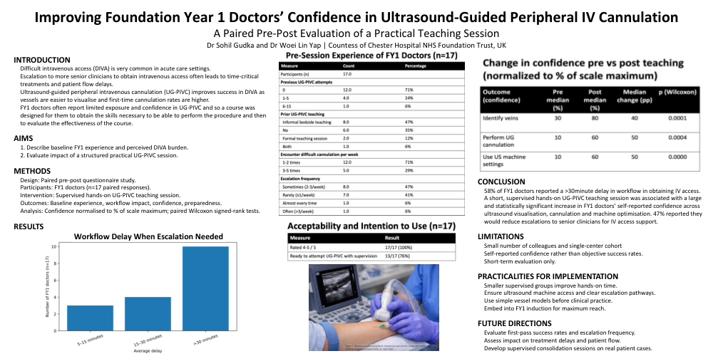
1. Describe baseline FY1 experience and perceived DIVA burden.
2. Evaluate impact of a structured practical UG-PIVC session.
METHODS
Design: Paired pre–post questionnaire study.
Participants: FY1 doctors (n=17 paired responses).
Intervention: Supervised hands-on UG-PIVC teaching session.
Outcomes: Baseline experience, workflow impact, confidence, preparedness.
Analysis: Confidence normalisedto % of scale maximum; paired Wilcoxon signed-rank tests.
CONCLUSION
58% of FY1 doctors reported a >30minute delay in workflow in obtaining IV access. A short, supervised hands-on UG-PIVC teaching session was associated with a large and statistically significant increase in FY1 doctors’ self-reported confidence across ultrasound visualisation, cannulation and machine optimisation. 47% reported they would reduce escalations to senior clinicians for IV access support.
LIMITATIONS
Small number of colleagues and single-center cohort
Self-reported confidence rather than objective success rates.
Short-term evaluation only.
PRACTICALITIES FOR IMPLEMENTATION
Smaller supervised groups improve hands-on time.
Ensure ultrasound machine access and clear escalation pathways.
Use simple vessel models before clinical practice.
Embed into FY1 induction for maximum reach.
FUTURE DIRECTIONS
Evaluate first-pass success rates and escalation frequency.
Assess impact on treatment delays and patient flow.
Develop supervised consolidation sessions on real patient cases.