140 posters, 10 videos, 15 audios, 20 sessions, 508 authors, 204 institutions
ePostersLive by SciGen Technologies S.A. All rights reserved.
8th World Congress on Vascular Access 2024
April 17 - 19, 2024 | Prague, Czech Republic

97
Venous port implantation failure due to inappropriate anatomical conditions - case reports of three oncology patients
Part of Topic
Introduction
Ultrasound guided port implantation and I-ECG navigation are commonly used techniques, enabling quick and safe procedure. However, inappropriate anatomical conditions are sometimes present, which makes ultrasound examination less clear and unrecognized venous anomalies can complicate an otherwise routine insertion.
Methods
We describe 3 case reports of patients with port implantation failure on the selected side. The first case concerned a female patient with biliary tract cancer, the two other patients had breast cancer. Despite the repeated successful puncture of the jugular vein (IJV), it was not possible to freely luminize the wire. CT angiography was indicated.
Results
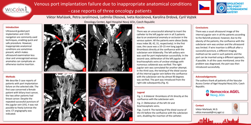
There was an unsuccessful attempt to insert the catheter to the left jugular vein in all 3 patients due to an anatomical anomaly or occlusion in the venous system. All the patients were obese (body mass index 38, 42, 31, respectively). In the first case, the cause was a 10–15 mm long jugular thrombosis directly at the confluence with the subclavian vein bilaterally. The left axillary vein was used for port implantation successfully. In the second case, obliteration of the left jugular and brachiocephalic veins of unclear etiology with numerous collaterals was verified. The right jugular vein was cannulated for another attempt. In the third case, the twisting of the distal course of the internal jugular vein before the confluence with the subclavian vein by almost 90 degrees was verified. The port was introduced from the right side subsequently (Fig. 4).
Conclusions
There was a usual ultrasound image of the internal jugular vein in all the patients according to the RaCeVA protocol, however, due to the obesity of the patients, the confluence with the subclavian vein was unclear and the ultrasound was limited. If wire insertion is difficult after a successful puncture, a different imaging method can be used in such patients and the port can be inserted using a different approach, if possible. In all the cases mentioned, once the problem was diagnosed, the port was then introduced successfully.