140 posters, 10 videos, 15 audios, 20 sessions, 508 authors, 204 institutions
ePostersLive by SciGen Technologies S.A. All rights reserved.
8th World Congress on Vascular Access 2024
April 17 - 19, 2024 | Prague, Czech Republic

95
Mechanisms and clinical presentations of the catheter injury during and after implantation of the totally implantable venous access port
Poster Presenter
Part of Topic
Background and objective: Catheter injury of the totally implantable venous access port (TIVAP) can lead to complications such as dysfunction or leakage of anticancer drugs. Sometimes leakage is a serious complication involving extensive necrosis of the tissue. It is important to be aware of the causes and mechanisms of catheter injury and make efforts to prevent it during the procedure. We present the mechanisms and clinical presentations of the catheter injury of TIVAP in our educational exhibition that includes radiographs and device photos.
MEHTODS: We retrospectively reviewed 14 cases referred for TIVAP removal due to complications related to catheter injury over the past 18 years. Medical records and radiographic images were evaluated for clinical presentation, presumed mechanism of injury, and management. If the catheter was not completely broken, cathetograms were obtained to assess the mechanism.
RESULTS: The clinical presentations of the catheter injury were complete breakage on radiographic image (n=7), leakage (n=4), and dysfunction (n=3). The most common presumed mechanism leading injury was pinch-off of the catheter (n=10) with access through the muscles (pectoralis, subclavius and sternocleidomastoid muscles). Other mechanisms included clamping of the catheter with Kelly during implantation (n=2), catheter puncture during suturing of the venipuncture site, (n=1), and injury during connection of the catheter to the reservoir (n=1). All catheters were successfully removed via femoral or axillary venous access after capture with a goose-neck snare.
CONCLUSIONS: Catheter injury in TIVAP can occur during and after the procedure due to various mechanisms and can lead to serious complications. For patency of TIVAP and patient safety, the implantation procedure should be performed with care to prevent catheter injury.
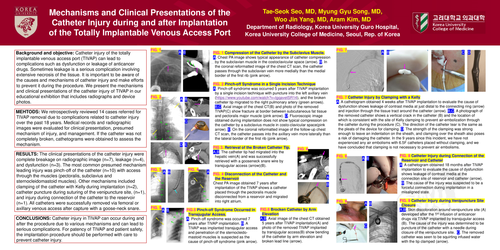
Compression of the Catheter by the Subclavius Muscle.
Chest PA image shows typical appearance of catheter compression by the subclavian muscle in the costoclavicular space. In the coronal reformatted image of the chest CT scan, the catheter passes through the subclavian vein more medially than the medial border of the first rib.
Pinch-off Syndrome in a Single Incision Technique
Pinch-off syndrome was occurred 5 years after TIVAP implantation by a single incision technique with puncture into the left axillary vein, and the broken catheter tip migrated to the right pulmonary artery (green arrows). Axial image of the chest CT and photo of the removed TIVAP show fracture at border between subcutaneous fat tissue and pectoralis major muscle. Fluoroscopic image obtained during implantation does not show typical compression on the catheter by a subclavius muscle in costoclavicular space. On the coronal reformatted image of the follow-up chest CT scan, the catheter passes into the axillary vein more laterally than the lateral border of the first rib.
Retrieval of the Broken Catheter Tip
The catheter tip had migrated into the hepatic vein and was successfully retrieved with a gooseneck snare wire by transjugular access.
Disconnection of the Catheter and the Reservoir
Chest PA image obtained 7 years after implantation of the TIVAP shows a catheter placed through the pectoralis muscle disconnected from a reservoir and migrated into right atrium.
Pinch-off Syndrome Occurred in Transjugular Access.
Pinch-off syndrome was occurred 7 years after TIVAP implantation. A TIVAP was implanted transjugular access and penetration of the sternocleidomastoid muscles is suspected as the cause of pinch-off syndrome.
Broken Catheter by Arm Elevation
Axial image of the chest CT obtained 5 years after TIVAP implantation and photo of the removed TIVAP implanted by transjugular access show bending of the catheter by arm elevation and broken lead line.
Catheter Injury by Clamping with a Kelly
A cathetogram obtained 4 weeks after TIVAP implantation to evaluate the cause of dysfunction shows leakage of contrast media at just distal to the connecting ring and injection through the tissue tract around the catheter. A photograph of the removed catheter shows a vertical crack in the catheter and the location of which is consistent with the site of Kelly clamping to prevent air-embolization through the catheter during the procedure. The direction of the catheter tear is the same as the pleats of the device for clamping. The strength of the clamping was strong enough to leave an indentation on the sheath, and clamping over the sheath also poses a risk of damaging the catheter. In the 9 years since this incident, we have not experienced any air embolisms with 6.5F catheters placed without clamping, and we have concluded that clamping is not necessary to prevent air embolisms.
Catheter Injury during Connection of the Reservoir and Catheter
A cathetogram obtained 18 months after TIVAP implantation to evaluate the cause of dysfunction shows leakage of contrast media at the connection site of reservoir and catheter. The cause of the injury was suspected to be a forceful connection during implantation in a misaligned state.
Catheter Injury during Venipuncture Site Closure
Skin discoloration around venipuncture site developed after the 1st infusion of anticancer drugs via TIVAP implanted by transjugular access. The cause of the injury was determined to be puncture of the catheter with a needle during closure of the venipuncture site. The removed catheter was seen to be squirting infused water with the tip clamped.