140 posters, 10 videos, 15 audios, 20 sessions, 508 authors, 204 institutions
ePostersLive by SciGen Technologies S.A. All rights reserved.
8th World Congress on Vascular Access 2024
April 17 - 19, 2024 | Prague, Czech Republic

183
Persistent left superior vena cava: accidental finding during the insertion of a peripherally inserted central catheter
Poster Presenter
Part of Topic
Audio
Persistent left superior vena cava: accidental finding during the insertion of a peripherally inserted central catheter
BACKGROUND
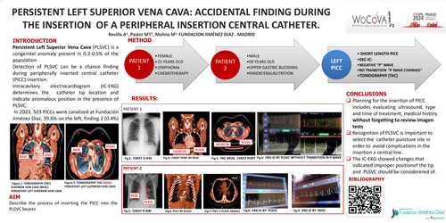
Persistent left superior vena cava (PLSVC) is a congenital anomaly present in 0.3-0.5% of the population. The 80-90% coexists with right superior vena cava (RSVC), 10-20% empty into the coronary sinus and 12% have heart disease. Detection of PLSVC may be a chance finding during peripherally inserted central catheter (PICC) insertion.
The intracavitary electrocardiogram (ECG-IC) indicates catheter tip location, by observing the variations in the “p” wave, which indicate improper position in the presence of PLSVC.
OBJETIVE
Describe the process of inserting the PICC into the PLSVC bearer
METHODS
In 2023, 503 PICCs were canalized, 39.6% on the left, finding 2 (0.4%) cases of PLSVC.
Two cases report:
21-year-old woman (P1), without medical history, diagnosed with Lymphoma with need for chemotherapy.
59-year-old male (P2), was hospitalized due to Upper Gastrointestinal Bleeding and was diagnosed with cancer, so central catheter was needed for parenteral nutrition.
Both patients were evaluated with ultrasound, finding in RaPeVa (Rapid Pheripheral Venous Assessment), a 55mm basilic vein on the left side and 40mm on the right side. PICC lines were canalized on the left in both patients. Under IC-ECG guidance we observed changes suggestive of abnormal position. Computed tomography (CT) confirmed the presence of PLSVC leading to the coronary sinus, coexisting with RSVC.
RESULTS
The estimated PICC measurement was 37cm in P1 and 44cm in P2, observing IC-ECG changes like premature negative “p waves”, without wave increase transition or maximum p wave, long before the estimated measurement.
Finally, catheters were canalized on the right side.
CONCLUSIONS
IC-ECG shows changes that indicate the improper tip location and the presence of PLSVC should be considered.
Recognition of PLSVC prior to insertion is important to select the catheter, puncture site and avoid complications.
Assessment of imaging tests is useful tool before PICC insertion