140 posters, 10 videos, 15 audios, 20 sessions, 508 authors, 204 institutions
ePostersLive by SciGen Technologies S.A. All rights reserved.
8th World Congress on Vascular Access 2024
April 17 - 19, 2024 | Prague, Czech Republic

166
Method to assess the feasibility of a novel ultrasound-based vein visualization device to assist with venous cannulation
Poster Presenter
Authors
Michael Liddelow, Ellaby Hansen, Phuoc Hao Ho, Katherine Arenson, Nicholas Buckley, Peter Carr, Barry Doyle, Nikhilesh Bappoo
Affiliations
VeinTech, VeinTech, School of Nursing and Midwifery, University of Galway, Vascular Engineering Laboratory, Harry Perkins Institute of Medical Research, QEII Medical Centre Nedlands and Centre for Medical Research, The University of Western Australia
Part of Topic
Video
Method to assess the feasibility of a novel ultrasound-based vein visualization device to assist with venous cannulation.
Cannulation is one of the most common invasive medical procedure (1); however, it is well established that standard manual techniques lead to an average 40% failure rate (2,3). Here we present a novel, ultraportable, ultrasound-based vein visualization device, and a simulation protocol to evaluate vein visualization and as a potential cannulation insertion training tool. This study aims to demonstrate the feasibility, repeatability, and reliability of the protocol and presents preliminary data on coronal view visualization usefulness.
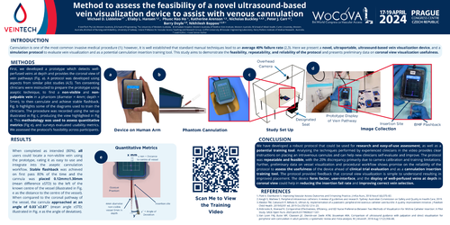
First, we developed a prototype which detects well-perfused veins at depth and provides the coronal view of vein pathways (Fig. a). A protocol was developed using aspects from similar pilot studies (4,5). Ten consenting clinicians were instructed to prepare the prototype using aseptic technique, to find a non-visible and non-palpable vein in a phantom (diameter = 4mm; depth = 5mm), to then cannulate and achieve stable flashback. Fig. b highlights some of the diagrams used to train the clinicians. The procedure was recorded using the set-up illustrated in Fig c, producing the view highlighted in Fig d. This methodology was used to assess quantitative metrics (Fig e), and surveys evaluated usability metrics. We assessed the protocol’s feasibility across participants.
When completed as intended (80%), all users could locate a non-visible vein using the prototype, rating it as easy to use and integrate into the aseptic cannulation workflow. Stable flashback was achieved on first pass 80% of the time and the cannula was placed 0.12mm±1.30mm (mean difference ±STD) to the left of the known centre of the vessel (illustrated in Fig. e as the distance to the centre of the vessel). When compared to the coronal pathway of the vessel, the cannula approached at an angle of0.03˚±2.87˚ (mean angle ±STD; illustrated in Fig. e as the angle of deviation).
We have developed a robust protocol that could be used for research and easy-of-use assessment, as well as a potential training tool. Analysing the techniques performed by experienced clinicians in the video provides clear instructions on placing an intravenous cannulas and can help new clinicians self-evaluate and improve. The protocol was repeatable and feasible, with the 20% discrepancy primarily due to camera calibration and training limitations. Further, preliminary data on vessel visualization and procedural workflow shows promise on the reliability of the protocol to assess theusefulness of the device ahead of clinical trial evaluation and as a cannulation insertion training tool. The protocol provided feedback that coronal view visualization is simple to understand resulting in improved placement. The device form factor, user-interface, and the display of well-perfused veins at depth in coronal view could help in reducing the insertion fail rate and improving correct vein selection.